. 2021 Mar 2;13(5):1056.
doi: 10.3390/cancers13051056.

Large Extracellular Vesicle Characterization and Association with Circulating Tumor Cells in Metastatic Castrate Resistant Prostate Cancer

Affiliations

Large Extracellular Vesicle Characterization and Association with Circulating Tumor Cells in Metastatic Castrate Resistant Prostate Cancer

Anna S Gerdtsson et al. Cancers (Basel). .

Abstract

Liquid biopsies hold potential as minimally invasive sources of tumor biomarkers for diagnosis, prognosis, therapy prediction or disease monitoring. We present an approach for parallel single-object identification of circulating tumor cells (CTCs) and tumor-derived large extracellular vesicles (LEVs) based on automated high-resolution immunofluorescence followed by downstream multiplexed protein profiling. Identification of LEVs >6 µm in size and CTC enumeration was highly correlated, with LEVs being 1.9 times as frequent as CTCs, and additional LEVs were identified in 73% of CTC-negative liquid biopsy samples from metastatic castrate resistant prostate cancer. Imaging mass cytometry (IMC) revealed that 49% of cytokeratin (CK)-positive LEVs and CTCs were EpCAM-negative, while frequently carrying prostate cancer tumor markers including AR, PSA, and PSMA. HSPD1 was shown to be a specific biomarker for tumor derived circulating cells and LEVs. CTCs and LEVs could be discriminated based on size, morphology, DNA load and protein score but not by protein signatures. Protein profiles were overall heterogeneous, and clusters could be identified across object classes. Parallel analysis of CTCs and LEVs confers increased sensitivity for liquid biopsies and expanded specificity with downstream characterization. Combined, it raises the possibility of a more comprehensive assessment of the disease state for precise diagnosis and monitoring.

Keywords: CTCs; aggressive variant prostate cancer; large extracellular vesicles; liquid biopsy; metastatic castrate-resistant prostate cancer; oncosomes.


Conflict of interest statement

James Hicks, Clinical Advisory Board, Epic Sciences, Inc. P. Kuhn received stock and dividends, Epic Sciences, Inc. Epic Sciences is evaluating the feasibility of translating the circulating tumor cell (CTC) assay into a commercial format.

Figures

Figure 1
Figure 1
Identification and size determination of circulating tumor cells and LEVs. (A) The integrated LEV/HDSCA workflow. (B) Examples of objects identified in one patient, classified to different categories and verified with higher resolution (40×) immunofluorescence and brightfield imaging. Composite shows DAPI in blue, pan-CK in red and CD45 in green. Scale bar is 10 µm. Arrows points to cell or LEV (DAPI-negative) for clarification. (C) Size distribution of LEVs, CTCs and apoptotic-like cells. (D) Linear regression of LEVs and CTCs with number of objects identified per slide, averaged over 2 slides analyzed per sample. (E) Cell/LEV enumeration grouped by tumor load (left panel) and bone marrow status (right panel). Open circles represent individual datapoints. Filled black circles and black line represent mean values and standard deviations, respectively.
Figure 1
Figure 1
Identification and size determination of circulating tumor cells and LEVs. (A) The integrated LEV/HDSCA workflow. (B) Examples of objects identified in one patient, classified to different categories and verified with higher resolution (40×) immunofluorescence and brightfield imaging. Composite shows DAPI in blue, pan-CK in red and CD45 in green. Scale bar is 10 µm. Arrows points to cell or LEV (DAPI-negative) for clarification. (C) Size distribution of LEVs, CTCs and apoptotic-like cells. (D) Linear regression of LEVs and CTCs with number of objects identified per slide, averaged over 2 slides analyzed per sample. (E) Cell/LEV enumeration grouped by tumor load (left panel) and bone marrow status (right panel). Open circles represent individual datapoints. Filled black circles and black line represent mean values and standard deviations, respectively.
Figure 2
Figure 2
Multiplex protein profiling of CTCs and LEVs. (A,B) Example of IF (top panels) and IMC (bottom panels) images of a CTC (A) and a LEV (B). The arrow points at the LEV which is negative for DAPI and weakly stained by the IMC DNA intercalator. Scale bar is 10 µm. (C) Heatmap of IMC scores on a 0–3 scale with 0 = white and dark blue = 3, ordered by object categories and patients, and clustered by proteins.
Figure 3
Figure 3
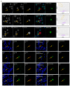
Biomarker expression in CTCs and LEVs of AVPC. (A) Example of HSPD1 expression in LEV (top panel), apoptotic (middle panel) and CTC (bottom panel). Cropped ROI images are shown with individual IF channels and IF composite image with DAPI (blue), CD45 (green) and pan-CK (red). The IMC rendered image of HSPD1 signal is shown in green. Table 1 vs. CD45 for all objects within the full ROI with WBCs in purple and the cell of interest (circled) in red (full cell), green (cytoplasm) and teal (nucleus). (B) Selected biomarkers shown for LEVs (top 2 panels) and CTCs (bottom 2 panels), with EpCAM expression varying from negative (0) to strong (3+). Scores from the 0–3 scale is shown for each marker and object. Scale bar is 10 µm.
Figure 4
Figure 4
Concurrent expression of EMT and epithelial markers detected by IMC analysis. (A) Subset of objects positive for EMT markers Twist-1 and Vimentin, most with concurrent expression of epithelial markers (EpCAM, CK8/18, E-cadherin). Selected individual channels from the CTC marked in (A) is shown in (B). In addition to the channels shown, this particular cell scored positive for HSPD1 (3+), HSPA5 (2+), ATP5B (1+), PD1 (1+) and CD24 (1+). CD31 (negative) was included to support that Vimentin-expression cannot be explained by endothelial origin. Scale bar is 10 µm.

References

    1. Hoshino A., Costa-Silva B., Shen T.L., Rodrigues G., Hashimoto A., Tesic Mark M., Molina H., Kohsaka S., Di Giannatale A., Ceder S., et al. Tumour exosome integrins determine organotropic metastasis. Nature. 2015;527:329–335. doi: 10.1038/nature15756. - DOI - PMC - PubMed
    1. Wu Z., Zhang Z., Xia W., Cai J., Li Y., Wu S. Extracellular vesicles in urologic malignancies-Implementations for future cancer care. Cell Prolif. 2019;52:e12659. doi: 10.1111/cpr.12659. - DOI - PMC - PubMed
    1. Minciacchi V.R., Spinelli C., Reis-Sobreiro M., Cavallini L., You S., Zandian M., Li X., Mishra R., Chiarugi P., Adam R.M., et al. MYC Mediates Large Oncosome-Induced Fibroblast Reprogramming in Prostate Cancer. Cancer Res. 2017;77:2306–2317. doi: 10.1158/0008-5472.CAN-16-2942. - DOI - PubMed
    1. Zanetti-Domingues L.C., Bonner S.E., Iyer R.S., Martin-Fernandez M.L., Huber V. Cooperation and Interplay between EGFR Signalling and Extracellular Vesicle Biogenesis in Cancer. Cells. 2020;9:2639. doi: 10.3390/cells9122639. - DOI - PMC - PubMed
    1. Murali V.P., Holmes C.A. Biomaterial-Based Extracellular Vesicle Delivery for Therapeutic Applications. Acta Biomater. 2021 doi: 10.1016/j.actbio.2021.01.010. - DOI - PubMed