. 2021 Mar 2;22(5):2499.
doi: 10.3390/ijms22052499.

Evaluation of Allogeneic Bone-Marrow-Derived and Umbilical Cord Blood-Derived Mesenchymal Stem Cells to Prevent the Development of Osteoarthritis in An Equine Model

Affiliations

Evaluation of Allogeneic Bone-Marrow-Derived and Umbilical Cord Blood-Derived Mesenchymal Stem Cells to Prevent the Development of Osteoarthritis in An Equine Model

Lélia Bertoni et al. Int J Mol Sci. .

Abstract

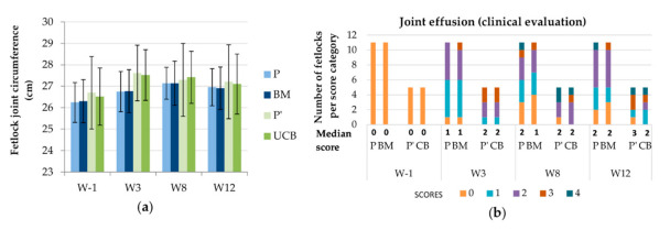
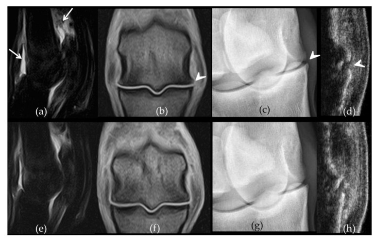
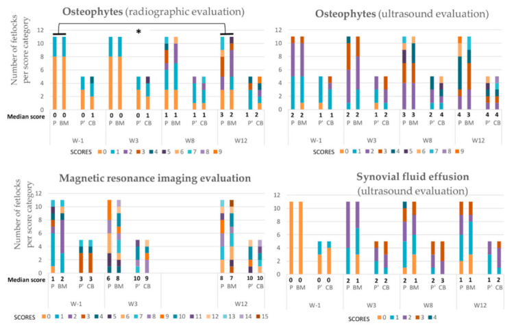
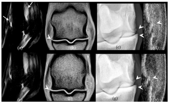
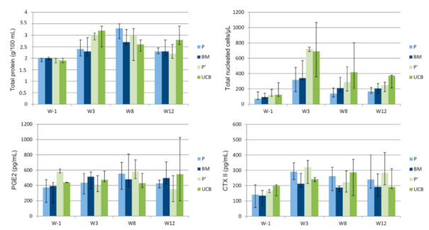
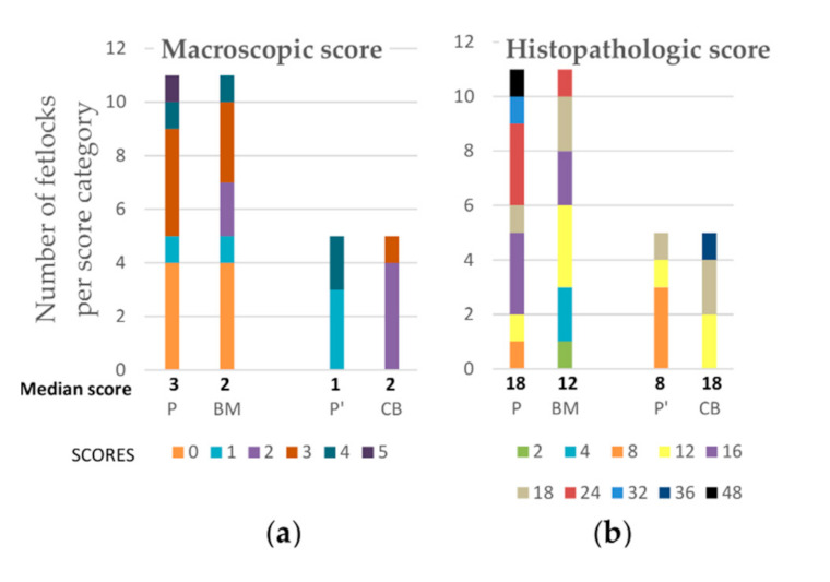
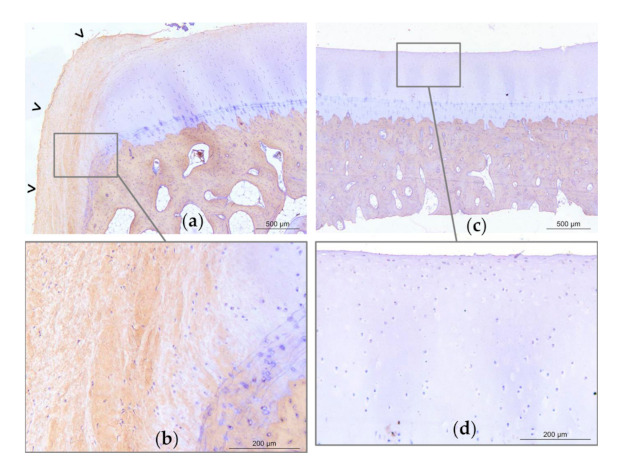
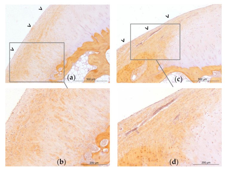
Osteoarthritis (OA) is a significant cause of pain in both humans and horses with a high socio-economic impact. The horse is recognized as a pertinent model for human OA. In both species, regenerative therapy with allogeneic mesenchymal stem cells (MSCs) appears to be a promising treatment but, to date, no in vivo studies have attempted to compare the effects of different cell sources on the same individuals. The objective of this study is to evaluate the ability of a single blinded intra-articular injection of allogeneic bone-marrow (BM) derived MSCs and umbilical cord blood (UCB) derived MSC to limit the development of OA-associated pathological changes compared to placebo in a post-traumatic OA model applied to all four fetlock joints of eight horses. The effect of the tissue source (BM vs. UCB) is also assessed on the same individuals. Observations were carried out using clinical, radiographic, ultrasonographic, and magnetic resonance imaging methods as well as biochemical analysis of synovial fluid and postmortem microscopic and macroscopic evaluations of the joints until Week 12. A significant reduction in the progression of OA-associated changes measured with imaging techniques, especially radiography, was observed after injection of bone-marrow derived mesenchymal stem cells (BM-MSCs) compared to contralateral placebo injections. These results indicate that allogeneic BM-MSCs are a promising treatment for OA in horses and reinforce the importance of continuing research to validate these results and find innovative strategies that will optimize the therapeutic potential of these cells. However, they should be considered with caution given the low number of units per group.

Keywords: allogeneic; bone marrow; horse; mesenchymal stem cells; osteoarthritis; pre-clinical study; umbilical cord blood.


Conflict of interest statement

The authors declare no conflict of interest.

Figures

Figure 1
Figure 1
A schematic presentation of the timeline of the study. JA—Joint assessment; Rx—Radiographic examination; Us—Ultrasound examination; MRI—Magnetic resonance imaging; SF—Synovial fluid sampling and analysis; Post-mortem—Post-mortem analysis including macroscopic and microscopic examination of the joints after euthanasia.
Figure 2
Figure 2
Evolution of two clinical parameters throughout the study period displayed per treatment group and per time point. (a) Mean (standard deviation) of the fetlock joint circumference. (b) Median scores and number of fetlocks per joint effusion score. W—Week; P—Fetlocks injected with placebo, contralateral of the fetlocks injected with bone-marrow derived mesenchymal stem cells (BM-MSCs); BM—Fetlocks injected with BM-MSCs; P’—Fetlocks injected with placebo, contralateral of the fetlocks injected with umbilical cord blood derived mesenchymal stem cells (UCB-MSCs); CB—Fetlocks injected with UCB-MSCs.
Figure 3
Figure 3
Comparative imaging findings observed on week 12 on the front metacarpophalangeal joints of horse 7. The left fore fetlock (ad) received placebo treatment while the right fore fetlock (eh) received bone-marrow derived mesenchymal stem cells (BM-MSCs) on week 3. On the left fetlock there was grade 3 (white arrows) synovial effusion (a) and periarticular osteophytes (b) (arrow heads) on Short Tau Inversion Recovery sagittal magnetic resonance imaging (MRI) images (a) and T1-weighed dorsal MRI images (b) respectively while grade 2 synovial effusion was observed on the right fetlock (ef). Similarly, there are grade 3 (arrow head) osteophytes on the dorsomedial-palmarolateral 35° oblique radiographic view from the left fore fetlock (c) compared to a grade 1 on the right (g). The ultrasound osteophyte score was 3 on the left (d) (arrow head) and 2 on the right fetlock (h), on the longitudinal sections made on the dorso-lateral aspect of both joints.
Figure 4
Figure 4
Evolution of the median scores and number of fetlocks per score category obtained for imaging parameters over the study period displayed per treatment group and per time point. W—Week; P—Fetlocks injected with placebo, contralateral of the fetlocks injected with bone-marrow derived mesenchymal stem cells (BM-MSCs); BM—Fetlocks injected with BM-MSCs; P’—Fetlocks injected with placebo, contralateral of the fetlocks injected with umbilical cord blood derived mesenchymal stem cells (UCB-MSCs); CB—Fetlocks injected with UCB-MSCs. * Significant difference between paired placebo and mesenchymal stem cell (MSC) treated fetlocks, considering the differences between W12 and W-1 values with p < 0.05.
Figure 5
Figure 5
Comparative imaging findings observed on week 12 on the front metacarpophalangeal joints of horse 4. The left fore fetlock (ad) received placebo treatment while the right fore fetlock (eh) received umbilical cord blood derived mesenchymal stem cells (UCB-MSCs)on week 3. On the left fetlock there was grade 3 (white arrows) synovial effusion (a) and periarticular osteophytes (b) (arrow heads) on Short Tau Inversion Recovery sagittal magnetic resonance imaging (MRI) images (a) and T1-weighed dorsal MRI images (b) respectively, while grades 2 were observed on the right fetlock (ef). There are grade 2 (arrow heads) osteophytes on the dorsomedial-palmarolateral 35° oblique radiographic views from both fore fetlocks (c,g) and on the dorso-lateral aspect of both joints on ultrasound (d,h) (arrow heads).
Figure 6
Figure 6
Evolution throughout the study period of the median values (1st quartile- 3rd quartile) of synovial fluid parameters displayed per treatment group. W—Week; P—Fetlocks injected with placebo, contralateral of the fetlocks injected with bone-marrow derived mesenchymal stem cells (BM-MSCs); BM—Fetlocks injected with BM-MSCs; P’—Fetlocks injected with placebo, contralateral of the fetlocks injected with umbilical cord blood derived mesenchymal stem cells (UCB-MSCs); UCB—Fetlocks injected with UCB-MSCs.
Figure 7
Figure 7
Median post-mortem scores and number of fetlocks per macroscopic (a) and histopathological (b) scores obtained on week 12 and displayed per treatment group. P—Fetlocks injected with placebo, contralateral of the fetlocks injected with bone-marrow derived mesenchymal stem cells (BM-MSCs); BM—Fetlocks injected with BM-MSCs; P’—Fetlocks injected with placebo, contralateral of the fetlocks injected with umbilical cord blood derived mesenchymal stem cells (UCB-MSCs); CB—Fetlocks injected with UCB-MSCs.
Figure 8
Figure 8
Representative light micrographs of osteochondral sections of the medial part of the distal aspect of the metacarpal condyles of horse 8. Micrographs obtained on Week 12, revealing type I collagen labelling (brown marking), and illustrating a grade 24 histological score on the left metacarpal condyle treated with placebo (ab) and a grade 4 on the right metacarpal condyle treated with bone-marrow derived mesenchymal stem cells (BM-MSCs) (cd). On the placebo treated joint (a), there is significant erosion of the superficial layer of the articular cartilage with a large fibrocellular tissue (open arrow heads) that is partly positive for type I collagen. On one extremity of the sample, cartilage and tidemark are no longer present (a). The subchondral bone appears trabecular. On the contralateral BM-MSC treated joint (c), the superficial layer of the cartilage is mildly eroded (d). There is no fibrocellular tissue, the tidemark is visible, and the subchondral bone is compact (c).
Figure 9
Figure 9
Representative light micrographs of osteochondral sections of the medial part of the distal aspect of the metacarpal condyles of horse 1. Micrographs obtained on Week 12, revealing type I collagen labelling (brown marking), and illustrating a grade 18 histological score on the left metacarpal condyle treated with umbilical cord blood derived mesenchymal stem cells (UCB-MSCs) (ab) and on the right metacarpal condyle treated with placebo (cd). On the UCB-MSC treated joint, there is mild erosion of the superficial layer of the articular cartilage (a). On both joints, there is a large fibrocellular tissue (open arrow heads) that is partly positive for type I collagen, and under which the tidemark is no longer present. The chondrocytes of the deep layers are also marked with type I collagen. The subchondral bone appears trabecular.

References

    1. Le Pen C., Reygrobellet C., Gérentes I. Les Conséquences Socioéconomiques de l’arthrose En France. Étude COART 1 France. Rev. Du Rhum. 2005;72:1326–1330. doi: 10.1016/j.rhum.2005.01.016. - DOI
    1. Lawrence R.C., Helmick C.G., Arnett F.C., Deyo R.A., Felson D.T., Giannini E.H., Heyse S.P., Hirsch R., Hochberg M.C., Hunder G.G. Estimates of the Prevalence of Arthritis and Selected Musculoskeletal Disorders in the United States. Arthritis Rheum. Off. J. Am. Coll. Rheumatol. 1998;41:778–799. doi: 10.1002/1529-0131(199805)41:5<778::AID-ART4>3.0.CO;2-V. - DOI - PubMed
    1. Oke S.L., McIlwraith C.W. Review of the Economic Impact of Osteoarthritis and Oral Joint-Health Supplements in Horses. Proc. Am. Assoc. Equine Pract. 2010;56:12–16.
    1. Shah K., Zhao A.G., Sumer H. New Approaches to Treat Osteoarthritis with Mesenchymal Stem Cells. Stem Cells Int. 2018;2018:5373294. doi: 10.1155/2018/5373294. - DOI - PMC - PubMed
    1. Neundorf R.H., Lowerison M.B., Cruz A.M., Thomason J.J., McEwen B.J., Hurtig M.B. Determination of the Prevalence and Severity of Metacarpophalangeal Joint Osteoarthritis in Thoroughbred Racehorses via Quantitative Macroscopic Evaluation. Am. J. Vet. Res. 2010;71:1284–1293. doi: 10.2460/ajvr.71.11.1284. - DOI - PubMed

Publication types

LinkOut - more resources