. 2021 Mar 2;10(3):530.
doi: 10.3390/cells10030530.

SARS-CoV-2 Nucleocapsid Protein Targets RIG-I-Like Receptor Pathways to Inhibit the Induction of Interferon Response

Affiliations

SARS-CoV-2 Nucleocapsid Protein Targets RIG-I-Like Receptor Pathways to Inhibit the Induction of Interferon Response

Soo Jin Oh et al. Cells. .

Abstract

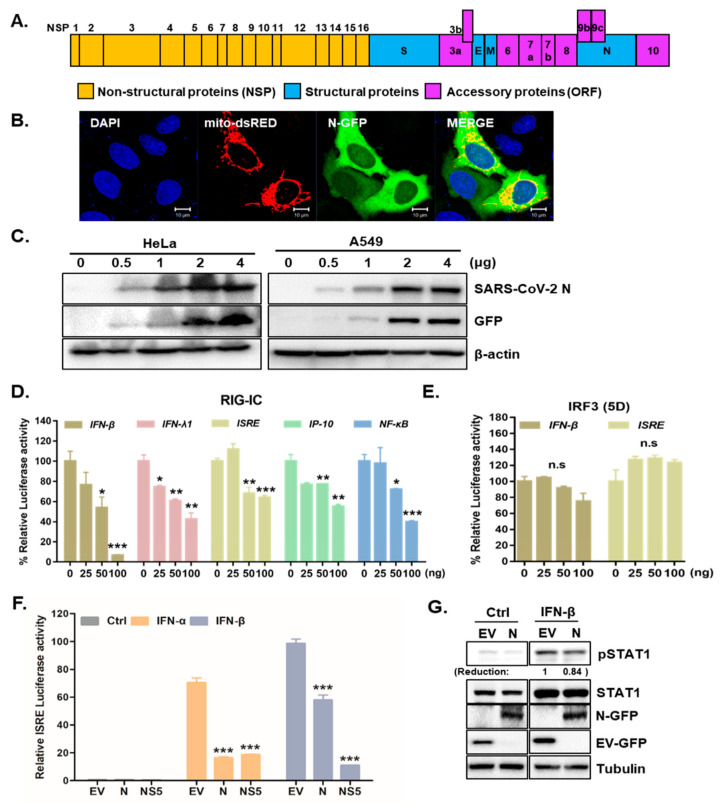
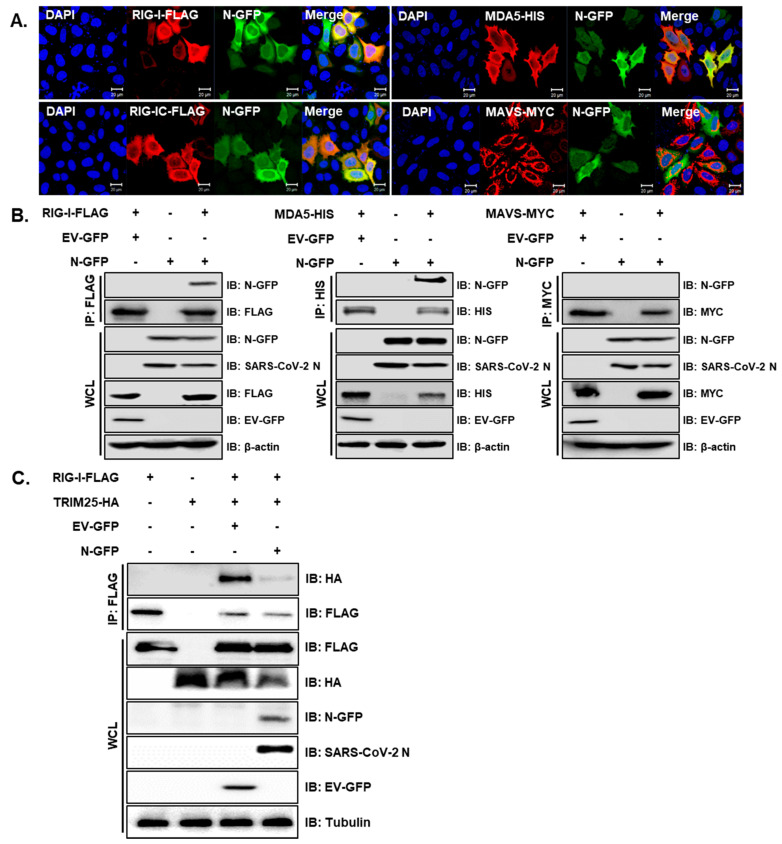
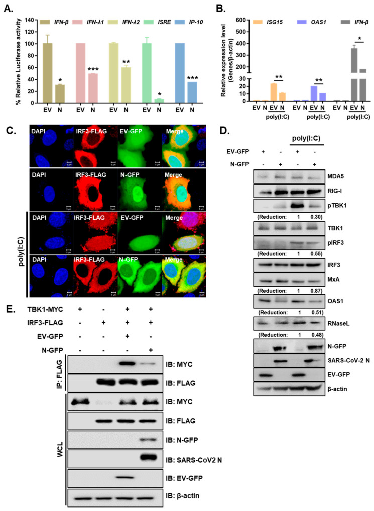
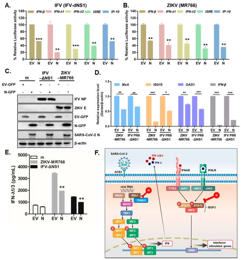
Severe acute respiratory syndrome coronavirus 2 (SARS-CoV-2) is the causative agent of the coronavirus disease 2019 (COVID-19) that has resulted in the current pandemic. The lack of highly efficacious antiviral drugs that can manage this ongoing global emergency gives urgency to establishing a comprehensive understanding of the molecular pathogenesis of SARS-CoV-2. We characterized the role of the nucleocapsid protein (N) of SARS-CoV-2 in modulating antiviral immunity. Overexpression of SARS-CoV-2 N resulted in the attenuation of retinoic acid inducible gene-I (RIG-I)-like receptor-mediated interferon (IFN) production and IFN-induced gene expression. Similar to the SARS-CoV-1 N protein, SARS-CoV-2 N suppressed the interaction between tripartate motif protein 25 (TRIM25) and RIG-I. Furthermore, SARS-CoV-2 N inhibited polyinosinic: polycytidylic acid [poly(I:C)]-mediated IFN signaling at the level of Tank-binding kinase 1 (TBK1) and interfered with the association between TBK1 and interferon regulatory factor 3 (IRF3), subsequently preventing the nuclear translocation of IRF3. We further found that both type I and III IFN production induced by either the influenza virus lacking the nonstructural protein 1 or the Zika virus were suppressed by the SARS-CoV-2 N protein. Our findings provide insights into the molecular function of the SARS-CoV-2 N protein with respect to counteracting the host antiviral immune response.

Keywords: RIG-I like receptors; SARS-CoV-2 N protein; antiviral immune response; coronavirus disease 2019; interferon.


Conflict of interest statement

The authors declare that the research was conducted in the absence of any commercial or financial relationships that could be construed as a potential conflict of interest.

Figures

Figure 1
Figure 1
SARS-CoV-2 N is a potent interferon antagonist. (A) Schematic view of SARS-CoV-2 genome is shown. (B) HeLa cells were transfected with SARS-CoV-2 N-GFP-encoding and mito-dsRED plasmids. Scale bar = 10 μm. (C) HeLa and A549 cells transfected with increasing amounts of SARS-CoV-2 N-GFP-encoding plasmids were immunoblotted with with the indicated antibodies to determine transfection efficiency. (D,E) HEK293T cells expressing RIG-IC, or IRF3 (5D) were co-transfected with firefly luciferase reporter plasmids encoding IFN-β, IFN-λ1, ISRE, IP-10, and NF-κB promoter together with increasing amount of SARS-CoV-2 N. The EV-GFP plasmid was used to ensure that each sample contains the same amount of total DNA plasmids. * p < 0.05; ** p < 0.01; *** p < 0.001, non-significant (n.s) versus EV-GFP-transfected cells (F) Recombinant IFN-α (50 ng/mL) or IFN-β (50 ng/mL) protein was added to the cells following the transfection with 100 ng of EV-GFP (EV), SARS-CoV-2 N (N) or ZIKV NS5 (NS5) encoding plasmids. Mean ± SD of three technical replicates are shown. *** p < 0.001 versus EV-GFP-transfected cells. (G) Immunoblot results of IFN-β-induced phosphorylation of STAT1 are shown. The images are representative of three independent experiments. Numbers indicate quantitative densitometric analyses of blot using ImageJ software.
Figure 2
Figure 2
SARS-CoV-2 N interacts with genes involved in the RLR signaling pathway and interferes RIG-I interaction with TRIM25. (A) HeLa cells were transiently transfected with the following plasmids: RIG-I-FLAG, RIG-IC-FLAG, MDA5-HIS and MAVS-MYC along with SARS-CoV-2 N-GFP-encoding plasmids. Cells were fixed in paraformaldehyde and permeabilized with 0.1% Triton X-100. The images are representative of three independent experiments. Scale bar = 20 μm. (B) HEK293T cells in 6-well plates were transfected with RIG-I-FLAG or MAVS-MYC plasmids along with empty vector with GFP tag (EV-GFP) or SARS-CoV-2 N-GFP (N-GFP) plasmids. After 24 h, cells were lysed and immunoprecipitated with indicated antibodies. Whole cell lysates (WCL) and immunoprecipitates were analyzed by immunoblot analysis. (C) HEK293T cells transfected with plasmids encoding RIG-I-FLAG or TRIM25-HA along with EV-GFP or N-GFP-encoding plasmids were lysed and subjected to immunoprecipitation with anti-FLAG tag antibody. The cell lysates and immunoprecipitates were analyzed by immunoblot analysis using indicated antibodies.
Figure 3
Figure 3
SARS-CoV-2 N inhibits the activation of IFN production and signaling in response to poly I:C treatment. (A) Luciferase activities of A549 cells transfected with 100 ng of EV-GFP (EV) or SARS-CoV-2 N-GFP (N) plasmids along with IFN-β, IFN-λ1, IFN-λ2, ISRE, and IP-10 promoter luciferase plasmids for 24 h and treated with poly (I:C) (10 µg/mL) for 6 h (mean ± SD, n = 3). * p < 0.05; ** p < 0.01; *** p < 0.001 versus EV-GFP-transfected cells. (B) A549 cells expressing either EV or N were transfected with poly (I:C) (10 µg/mL) for 6 h. RT-qPCR analysis of ISG15, OAS1, and IFN-β mRNAs is shown (mean ± SD, n = 3). * p < 0.05; ** p < 0.01 versus EV-GFP-transfected cells.(C) A549 cells were co-transfected with IRF3-FLAG and EV-GFP or N-GFP plasmids. After 24 h, cells were treated with poly I:C (10 µg/mL) for 2 h and fixed to observe the subcellular localization of IRF3. Scale bar = 5 μm. (D) A549 cells were transiently transfected with EV-GFP or N-GFP plasmids. After 24 h, cells were transfected with poly (I:C) (10 µg/mL) for 2 h. Cell lysates were collected for immunoblotting with the indicated antibodies. Numbers indicate quantitative densitometric analyses of blot using ImageJ software. (E) HEK293T cells were transfected with plasmids encoding IRF3-FLAG and TBK1-MYC along with EV-GFP or N-GFP for 24 h. Cell lysates were subjected to immunoprecipitation with an anti-FLAG tag antibody, followed by immunoblot analysis with indicated antibodies.
Figure 4
Figure 4
SARS-CoV-2 N markedly attenuates RNA virus-triggered IFN production. (A,B) Luciferase activities of A549 cells transfected with IFN-β, IFN-λ1, IFN-λ2, ISRE, and IP-10 promoter luciferase plasmids together with expression vector for RIG-I, along with 100 ng of EV-GFP (EV) or SARS-CoV-2 N-GFP (N) expression plasmids. At 24 h after transfection, the cells were infected with IFV-delNS1 (IFV-dNS1) (A) or ZIKV (MR766) (B) at multiplicity of infection (MOI) = 1 for 6 h (mean ± SD, n = 3). ** p < 0.01; *** p < 0.001 versus EV-GFP-transfected cells. (C) A549 cells were transiently transfected with EV-GFP or SARS-CoV-2 N-GFP expression plasmids. After 24 h, cells were infected with mock (m), ZIKV (MR766), or IFV-delNS1 (MOI = 1). (D) RT-qPCR reveals mRNA levels of MxA, IFN-β, ISG15, and OAS1 following viral infection. * p < 0.05; ** p < 0.01; *** p < 0.001 versus EV-GFP-transfected cells. (E) IFN-λ1/3 secretion levels were measured by ELISA assays. ** p < 0.01 versus EV-GFP-transfected cells. (F) Schematic representation of how SARS-CoV-2 N protein interferes with IFN induction and signaling.

References

    1. Huang C., Wang Y., Li X., Ren L., Zhao J., Hu Y., Zhang L., Fan G., Xu J., Gu X., et al. Clinical features of patients infected with 2019 novel coronavirus in Wuhan, China. Lancet. 2020;395:497–506. doi: 10.1016/S0140-6736(20)30183-5. - DOI - PMC - PubMed
    1. Vabret N., Britton G.J., Gruber C., Hegde S., Kim J., Kuksin M., Levantovsky R., Malle L., Moreira A., Park M.D., et al. Immunology of COVID-19: Current State of the Science. Immunity. 2020;52:910–941. doi: 10.1016/j.immuni.2020.05.002. - DOI - PMC - PubMed
    1. Cui J., Li F., Shi Z.L. Origin and evolution of pathogenic coronaviruses. Nat. Rev. Microbiol. 2019;17:181–192. doi: 10.1038/s41579-018-0118-9. - DOI - PMC - PubMed
    1. Zhu N., Zhang D., Wang W., Li X., Yang B., Song J., Zhao X., Huang B., Shi W., Lu R., et al. A Novel Coronavirus from Patients with Pneumonia in China, 2019. N. Engl. J. Med. 2020;382:727–733. doi: 10.1056/NEJMoa2001017. - DOI - PMC - PubMed
    1. Schneider W.M., Chevillotte M.D., Rice C.M. Interferon-stimulated genes: A complex web of host defenses. Annu. Rev. Immunol. 2014;32:513–545. doi: 10.1146/annurev-immunol-032713-120231. - DOI - PMC - PubMed

Publication types

MeSH terms

LinkOut - more resources