. 2021 Mar 2;10(3):531.
doi: 10.3390/cells10030531.

Deficiency of Biogenic Amines Modulates the Activity of Hypoglossal Nerve in the Reserpine Model of Parkinson's Disease

Affiliations

Deficiency of Biogenic Amines Modulates the Activity of Hypoglossal Nerve in the Reserpine Model of Parkinson's Disease

Monika Jampolska et al. Cells. .

Abstract

The underlying cause of respiratory impairments appearing in Parkinson's disease (PD) is still far from being elucidated. To better understand the pathogenesis of respiratory disorders appearing in PD, we studied hypoglossal (HG) and phrenic (PHR) motoneuron dysfunction in a rat model evoked with reserpine administration. After reserpine, a decrease in the baseline amplitude and minute HG activity was noted, and no depressive phase of the hypoxic ventilatory response was observed. The pre-inspiratory time of HG activity along with the ratio of pre-inspiratory time to total respiratory cycle time and the ratio of pre-inspiratory to inspiratory amplitude were significantly reduced during normoxia, hypoxia, and recovery compared to sham rats. We suggest that the massive depletion of not only dopamine, but above all noradrenaline and serotonin in the brainstem observed in our study, has an impact on the pre-inspiratory activity of the HG. The shortening of the pre-inspiratory activity of the HG in the reserpine model may indicate a serious problem with maintaining the correct diameter of the upper airways in the preparation phase for inspiratory effort and explain the development of obstructive sleep apnea in some PD patients. Therapies involving the supplementation of amine depletion other than dopamine should be considered.

Keywords: Parkinson’s diseases; biogenic amines; hypoglossal nerve; hypoxia; phrenic nerve; reserpine model.


Conflict of interest statement

The authors declare no conflict of interest.

Figures

Figure 1
Figure 1
Sample record of neurograms of the hypoglossal nerve (HG, XII nerve) and the phrenic nerve (PHR) showing the calculation method of the following parameters: inspiratory time (TI), expiratory time (TE), pre-inspiratory time of HG nerve activity (T pre-I), the amplitude (A) of HG and PHR, amplitude of HG pre-inspiratory activity (A pre-I HG).
Figure 2
Figure 2
The average amplitude (A,B) and minute activity (A × f) of the hypoglossal nerve (HG) (C,D) during normoxia, hypoxic ventilatory response, and recovery in animals subjected to a vehicle (sham) or reserpine injection. Results are expressed as absolute values (A,C) and a percentage of the baseline nerve activity (expressed as 100%) (B,D) before hypoxia. All values are means ± SEM. *# P < 0.05, **## P < 0.01, ***### P < 0.001,*—statistical significance in comparison to normoxic value, #—statistical significance between corresponding values in reserpine and sham groups (n = 7–9 per group).
Figure 3
Figure 3
The changes in amplitude (A,B), minute activity (A × f) (C,D), and frequency of discharge (f) (E,F) of the phrenic nerve (PHR) during normoxia, hypoxic ventilatory response, and recovery in animals subjected to a vehicle (sham) or reserpine injection. Results are expressed as absolute values (A,C,E) and percentage of the baseline nerve activity (considered 100%) (B,D,F) before hypoxia. All values are means ± SEM. *# P < 0.05; **## P < 0.01, ***### P < 0.001, *—statistical significance in comparison to normoxic value, #—statistical significance between corresponding values in reserpine and sham groups (n = 7–9 per group).
Figure 4
Figure 4
The changes in time of inspiration (TI) (A), time of expiration (TE) (B), and the total time of respiratory cycle (TC) (C) in the respiratory response to hypoxia in rats injected i.p. with vehicle (sham) or reserpine. All values are given as mean ± SEM. *# P < 0.05; **## P < 0.01, *—statistical significance in comparison to normoxic value, #—statistical significance between corresponding values in reserpine and sham groups (n = 7–9 per group).
Figure 5
Figure 5
The average pre-inspiratory hypoglossal nerve amplitude (A pre-I HG) (A), a ratio of the pre-inspiratory hypoglossal nerve amplitude to the inspiratory HG peak amplitude (A pre-I HG/A HG) (B), pre-inspiratory time of HG (T pre-I) (C) and the ratio of pre-inspiratory time of HG to a total length of the respiratory cycle (T pre-I/Tc) (D) in respiratory response to hypoxia in rats injected i.p. with vehicle (sham) or reserpine. All values are given as mean ± SEM. All values are given as mean ± SEM. *# P < 0.05; ## P < 0.01, *—statistical significance in comparison to normoxic value, #—statistical significance between corresponding values in reserpine and sham groups (n = 7–9 per group).
Figure 6
Figure 6
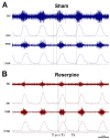
Typical neurogram records of phrenic (PHR) and hypoglossal (HG) nerve activity (raw and integrated signals—∫) in sham (A) and reserpine (B) rats during hypoxia. Note the apparently shortened time of inspiration (TI) and pre-inspiratory time of HG nerve activity (T pre-I) in the reserpine rats compared to the sham animals.

Similar articles

Cited by

References

    1. Hirsch E., Graybiel A.M., Agid Y.A. Melanized dopaminergic neurons are differentially susceptible to degeneration in Parkinson’s disease. Nature. 1988;334:345–348. doi: 10.1038/334345a0. - DOI - PubMed
    1. Stuart S., Morris R., Giritharan A., Quinn J., Nutt J.G., Mancini M. Prefrontal Cortex Activity and Gait in Parkinson’s Disease With Cholinergic and Dopaminergic Therapy. Mov. Disord. 2020;35:2019–2027. doi: 10.1002/mds.28214. - DOI - PubMed
    1. da Silva Córneo E., de Bem Silveira G., Scussel R., Borges Correa M.E.A., da Silva Abel J., Luiz G.P., Feuser P.E., Silveira P.C.L., Machado-de-Ávila R.A. Effects of gold nanoparticles administration through behavioral and oxidative parameters in animal model of Parkinson’s disease. Colloids Surf. B Biointerfaces. 2020;196:111302. doi: 10.1016/j.colsurfb.2020.111302. - DOI - PubMed
    1. Baille G., De Jesus A.M., Perez T., Devos D., Dujardin K., Charley C.M., Defebvre L., Moreau C. Ventilatory Dysfunction in Parkinson’s Disease. J. Parkinsons Dis. 2016;6:463–471. doi: 10.3233/JPD-160804. - DOI - PMC - PubMed
    1. Pokusa M., Hajduchova D., Buday T., Kralova Trancikova A. Respiratory Function and Dysfunction in Parkinson-Type Neurodegeneration. Physiol. Res. 2020;69:S69–S79. doi: 10.33549/physiolres.934405. - DOI - PMC - PubMed

LinkOut - more resources