Review
. 2021 Mar 27;13(7):1547.
doi: 10.3390/cancers13071547.

Pineal Gland Tumors: A Review

Affiliations
Review

Pineal Gland Tumors: A Review

Gaia Favero et al. Cancers (Basel). .

Abstract

The pineal gland is a small, pinecone-shaped endocrine gland that participates in the biological rhythm regulation of vertebrates. The recognized major product of the pineal gland is melatonin-a multifunctional endogenous indoleamine. Accumulating evidence suggests that the pineal gland is important for preserving ideal health conditions in vertebrate. Tumors of the pineal region account for approximately 3-11% of pediatric brain neoplasms but fewer than 1% of brain neoplasms in adults. It is fundamental to expand advanced imaging techniques together with both clinical and laboratory knowledge, to help to differentiate among pineal neoplasms and thus facilitate accurate primary diagnoses and proper therapeutic interventions. In this review, we report the gross anatomy of the pineal gland and its functional significance and discuss the clinical relevance of pineal gland tumors, underlining the importance of identifying the leading causes of pineal region masses.

Keywords: brain neoplasms; pineal germ cell tumors; pineal gland; pineal metastasis; pineal parenchymal tumor.


Conflict of interest statement

The authors declare no conflict of interest.

Figures

Figure 1
Figure 1
Human pineal gland (a) and its anatomic boundaries (b). The pineal gland is visible in yellow (a). Anatomage Inc.—Anatomage Table EDU. The 3D rendering of the cadaver data is from Anatomage Table.
Figure 2
Figure 2
Pineal tumor classification.
Figure 3
Figure 3
Pineal germinoma magnetic resonance imaging (MRI). Pineal tumor hypointense on T1 weighted image (T1WI) (A), hyperintense on T2WI (B) and with homogeneous contrast enhancement (C). The arrow identifies hyperdense mass with calcification at computed tomography (D). In another patient, a larger germinoma, isointense on T1WI (E). Reprinted with permission from Reis et al. (2006) [41]. John Wiley and Sons—2021 (License Number 5034711197310).
Figure 4
Figure 4
Pineal choriocarcinoma MRI. Susceptibility-weighted imaging (A), T2WI (B) and pineal heterogeneous mass on T1WI (C,D). The arrows identify the faint peripheral enhancement after the intravenous administration of gadolinium (E). Reprinted with permission from Causil et al. (2016) [46]. Elsevier—2021 (License Number: 5034730033295).
Figure 5
Figure 5
Pineal teratoma MRI. Sagittal T1 image (A), axial T1 (B) and sagittal contrast-enhanced T1 (C) images. Reprinted with permission from Peterson et al. (2012) [54]. Elsevier—2021 (License Number: 5034851378505).
Figure 6
Figure 6
Pineocytoma MRI. The asterisk indicates the homogeneous mass in the pineal gland. Reprinted with permission from Smirniotopoulos et al. (1992) [13]. Radiological Society North America–2021. Small pineocytomas often do not induce symptoms, but if they are of large dimensions, they may induce obstructive hydrocephalus and Parinaud syndrome, defined as upward gaze palsy, pupillary light-near dissociation, and convergence retraction nystagmus [15]. The five- and twenty-year survival rates are, respectively, 100% and 76% [21,26]. For pineocytomas, stereotactic radiosurgery seems to be highly effective as a primary treatment, so stereotactic radiosurgery alone may be considered appropriate clinical management for pineocytomas [21,28,71,72].
Figure 7
Figure 7
Pineoblastoma MRI. Preoperative MRI (AC), MRI 1.5 months after surgery showing spinal metastasis (D). Reprinted with permission from Huo et al. (2020) [79]. Dove Medical Press—2021.
Figure 8
Figure 8
Pineal papillary tumor MRI before (A) and after 15 years of treatments (B). Reprinted with permission from Fernández-Mateos et al. (2018) [90]. Elsevier—2021 (License Number: 5034870401490).
Figure 9
Figure 9
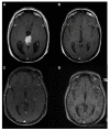
Pineal parenchymal tumors of intermediate differentiation MRI. Preoperative MRI (A), postoperative MRI showing a very small residual pineal lesion (B). MRI before (C) and after (D) radiation therapy. Reprinted with permission from Choque- Velasquez et al. (2019) [102]. Elsevier—2021 (License Number: 5035440109275). Pineal parenchymal tumors of intermediate differentiation may be morphologically classified into (1) lobulated endocrine-like and highly vascular lesions; (2) those with diffuse growth patterns, similar to oligodendrogliomaneurocytomas; and (3) those of transitional type, with areas of lobulated and diffuse growth patterns, correlated with areas of pineocytomatous rosettes [82,102,109]. Recently, Wu et al. [103] reported that CD24 and PRAME may be novel markers useful for the grading and prognostic evaluation of pineal parenchymal tumors of intermediate differentiation and, thus, useful in therapeutic decision-making. The best treatment for pineal parenchymal tumors of intermediate differentiation has not yet been found, partly due to the low numbers of reported cases. The maximal surgical removal of the tumor is the optimal treatment in practice for pineal parenchymal tumors of intermediate differentiation. However, even after complete surgical tumor removal, many patients experience recurrence. Hence, adjuvant radio- or chemo-therapy, or a mix of both, is often recommended to improve patient survival. A report from the International Gamma Knife Research Foundation described actuarial local control and survival rates following stereotactic radiosurgery on pineal parenchymal tumors of intermediate differentiation of 50% and 56%, respectively, at 5 years [19,26].
Figure 10
Figure 10
Pineal metastasis from lung adenocarcinoma. Brain MRI sagittal (A), coronal (B) and transverse (C) sections showing the pineal gland metastatic mass. Computed tomography showing a lung nodule over the left hilar region in the lower right corner (D). Image from Abdallah et al. (2020) [110]—Ochsner Journal.
Figure 11
Figure 11
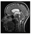
Pineal cyst MRI. The asterisk indicates the pineal cyst. Reprinted with permission from Májovský et al. (2017) [124]—Elsevier—2021 (License Number 5020240575766).

Similar articles

  • Biomarkers of Pineal Region Tumors: A Review.
    Carr C, O'Neill BE, Hochhalter CB, Strong MJ, Ware ML. Carr C, et al. Ochsner J. 2019 Spring;19(1):26-31. doi: 10.31486/toj.18.0110. Ochsner J. 2019. PMID: 30983898 Free PMC article. Review.
  • Pineal Gland Tumor but not Pinealoma: A Case Report.
    Naqvi S, Rupareliya C, Shams A, Hameed M, Mahuwala Z, Giyanwani PR. Naqvi S, et al. Cureus. 2017 Aug 18;9(8):e1576. doi: 10.7759/cureus.1576. Cureus. 2017. PMID: 29057188 Free PMC article.
  • Pineal Region Masses in Pediatric Patients.
    Tamrazi B, Nelson M, Blüml S. Tamrazi B, et al. Neuroimaging Clin N Am. 2017 Feb;27(1):85-97. doi: 10.1016/j.nic.2016.08.002. Neuroimaging Clin N Am. 2017. PMID: 27889025 Review.
  • MRI of pineal region tumors.
    Korogi Y, Takahashi M, Ushio Y. Korogi Y, et al. J Neurooncol. 2001 Sep;54(3):251-61. doi: 10.1023/a:1012773727022. J Neurooncol. 2001. PMID: 11767291 Review.
  • Pineal Gland Cancer.
    Mayol Del Valle M, De Jesus O. Mayol Del Valle M, et al. 2023 Aug 23. In: StatPearls [Internet]. Treasure Island (FL): StatPearls Publishing; 2025 Jan–. 2023 Aug 23. In: StatPearls [Internet]. Treasure Island (FL): StatPearls Publishing; 2025 Jan–. PMID: 32809402 Free Books & Documents.

Cited by

References

    1. Carr C., O’Neill B.E., Hochhalter C.B., Strong M.J., Ware M.L. Biomarkers of pineal region tumors: A review. Ochsner J. 2019;19:26–31. doi: 10.31486/toj.18.0110. - DOI - PMC - PubMed
    1. Fèvre-Montange M., Vasiljevic A., Champier J., Jouvet A. Histopathology of tumors of the pineal region. Future Oncol. 2010;6:791–809. doi: 10.2217/fon.10.28. - DOI - PubMed
    1. Nagasawa D.T., Lagman C., Sun M., Yew A., Chung L.K., Lee S.J., Bui T.T., Ooi Y.C., Robison R.A., Zada G., et al. Pineal germ cell tumors: Two cases with review of histopathologies and biomarkers. J. Clin. Neurosci. 2017;38:23–31. doi: 10.1016/j.jocn.2016.12.024. - DOI - PMC - PubMed
    1. Fernández-Palanca P., Méndez-Blanco C., Fondevila F., Tuñón M.J., Reiter R.J., Mauriz J.L., González-Gallego J. Melatonin as an antitumor agent against liver cancer: An updated systematic review. Antioxidants. 2021;10:103. doi: 10.3390/antiox10010103. - DOI - PMC - PubMed
    1. Moretti R., Zanin A., Pansiot J., Spiri D., Manganozzi L., Kratzer I., Favero G., Vasiljevic A., Rinaldi V.E., Pic I., et al. Melatonin reduces excitotoxic blood-brain barrier breakdown in neonatal rats. Neuroscience. 2015;311:382–397. doi: 10.1016/j.neuroscience.2015.10.044. Erratum in: Neuroscience2016, 315, 296. - DOI - PubMed

LinkOut - more resources