Soluble JAM-C Ectodomain Serves as the Niche for Adipose-Derived Stromal/Stem Cells
- PMID: 33801826
- PMCID: PMC8000331
- DOI: 10.3390/biomedicines9030278
Soluble JAM-C Ectodomain Serves as the Niche for Adipose-Derived Stromal/Stem Cells
Abstract
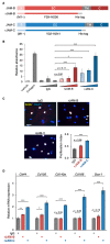
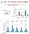
Junctional adhesion molecules (JAMs) are expressed in diverse types of stem and progenitor cells, but their physiological significance has yet to be established. Here, we report that JAMs exhibit a novel mode of interaction and biological activity in adipose-derived stromal/stem cells (ADSCs). Among the JAM family members, JAM-B and JAM-C were concentrated along the cell membranes of mouse ADSCs. JAM-C but not JAM-B was broadly distributed in the interstitial spaces of mouse adipose tissue. Interestingly, the JAM-C ectodomain was cleaved and secreted as a soluble form (sJAM-C) in vitro and in vivo, leading to deposition in the fat interstitial tissue. When ADSCs were grown in culture plates coated with sJAM-C, cell adhesion, cell proliferation and the expression of five mesenchymal stem cell markers, Cd44, Cd105, Cd140a, Cd166 and Sca-1, were significantly elevated. Moreover, immunoprecipitation assay showed that sJAM-C formed a complex with JAM-B. Using CRISPR/Cas9-based genome editing, we also demonstrated that sJAM-C was coupled with JAM-B to stimulate ADSC adhesion and maintenance. Together, these findings provide insight into the unique function of sJAM-C in ADSCs. We propose that JAMs contribute not only to cell-cell adhesion, but also to cell-matrix adhesion, by excising their ectodomain and functioning as a niche-like microenvironment for stem and progenitor cells.
Keywords: junctional adhesion molecule; mesenchymal stem cell; niche; shedding; stem cell; tight junction.
Conflict of interest statement
The authors declare no conflict of interest.
Figures


Similar articles
-
Junctional adhesion molecule-C is a soluble mediator of angiogenesis.J Immunol. 2010 Aug 1;185(3):1777-85. doi: 10.4049/jimmunol.1000556. Epub 2010 Jun 30. J Immunol. 2010. PMID: 20592283 Free PMC article.
-
Differential expression of junctional adhesion molecules in different stages of systemic sclerosis.Arthritis Rheum. 2013 Jan;65(1):247-57. doi: 10.1002/art.37712. Arthritis Rheum. 2013. PMID: 23001478
-
Junctional adhesion molecule A expressed on human CD34+ cells promotes adhesion on vascular wall and differentiation into endothelial progenitor cells.Arterioscler Thromb Vasc Biol. 2010 Jun;30(6):1127-36. doi: 10.1161/ATVBAHA.110.204370. Epub 2010 Apr 8. Arterioscler Thromb Vasc Biol. 2010. PMID: 20378847
-
Junctional Adhesion Molecules (JAMs): The JAM-Integrin Connection.Cells. 2018 Mar 26;7(4):25. doi: 10.3390/cells7040025. Cells. 2018. PMID: 29587442 Free PMC article. Review.
-
Junctional Adhesion Molecules in Cancer: A Paradigm for the Diverse Functions of Cell-Cell Interactions in Tumor Progression.Cancer Res. 2020 Nov 15;80(22):4878-4885. doi: 10.1158/0008-5472.CAN-20-1829. Epub 2020 Aug 14. Cancer Res. 2020. PMID: 32816855 Free PMC article. Review.
Cited by
-
Increased Circulating Soluble Junctional Adhesion Molecules in Systemic Sclerosis: Association with Peripheral Microvascular Impairment.Life (Basel). 2022 Nov 4;12(11):1790. doi: 10.3390/life12111790. Life (Basel). 2022. PMID: 36362943 Free PMC article.
-
Junctional adhesion molecule-C: A multifunctional mediator of cell adhesion.Cell Mol Life Sci. 2025 Aug 13;82(1):312. doi: 10.1007/s00018-025-05829-z. Cell Mol Life Sci. 2025. PMID: 40801950 Free PMC article. Review.
References
-
- Friedenstein A.J., Piatetzky-Shapiro I.I., Petrakova K.V. Osteogenesis in transplants of bone marrow cells. J. Embryol. Exp. Morphol. 1966;16:381–390. - PubMed
-
- Sacchetti B., Funari A., Michienzi S., Di Cesare S., Piersanti S., Saggio I., Tagliafico E., Ferrari S., Robey P.G., Riminucci M., et al. Self-renewing osteoprogenitors in bone marrow sinusoids can organize a hematopoietic microenvironment. Cell. 2007;131:324–336. doi: 10.1016/j.cell.2007.08.025. - DOI - PubMed
Grants and funding
LinkOut - more resources
Full Text Sources
Other Literature Sources
Research Materials
Miscellaneous

