. 2021 Mar 10;9(3):239.
doi: 10.3390/vaccines9030239.

Immunogenicity of HIV-1-Based Virus-Like Particles with Increased Incorporation and Stability of Membrane-Bound Env

Affiliations

Immunogenicity of HIV-1-Based Virus-Like Particles with Increased Incorporation and Stability of Membrane-Bound Env

Christopher A Gonelli et al. Vaccines (Basel). .

Abstract

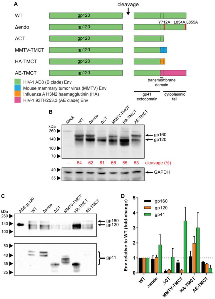
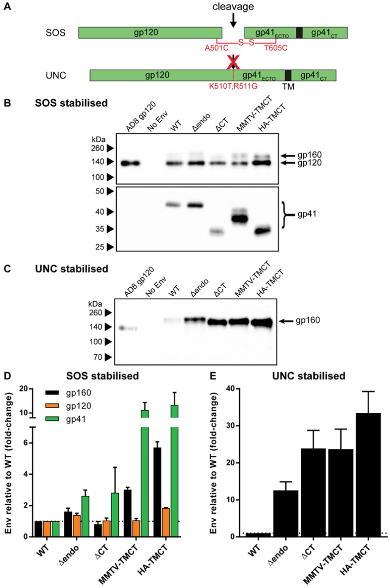
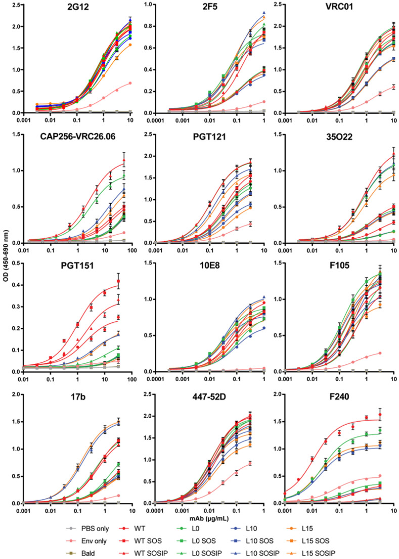
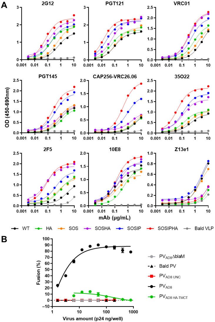
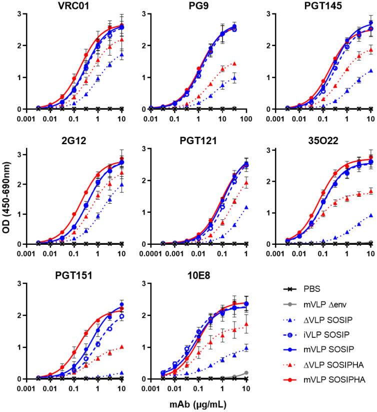
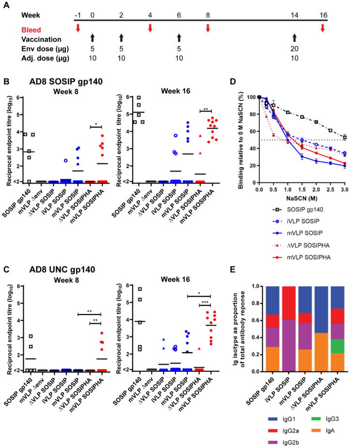
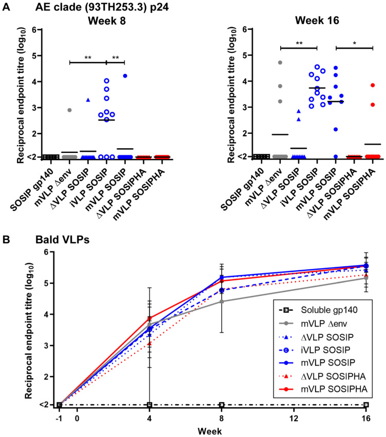
An optimal prophylactic vaccine to prevent human immunodeficiency virus (HIV-1) transmission should elicit protective antibody responses against the HIV-1 envelope glycoprotein (Env). Replication-incompetent HIV-1 virus-like particles (VLPs) offer the opportunity to present virion-associated Env with a native-like structure during vaccination that closely resembles that encountered on infectious virus. Here, we optimized the incorporation of Env into previously designed mature-form VLPs (mVLPs) and assessed their immunogenicity in mice. The incorporation of Env into mVLPs was increased by replacing the Env transmembrane and cytoplasmic tail domains with those of influenza haemagglutinin (HA-TMCT). Furthermore, Env was stabilized on the VLP surface by introducing an interchain disulfide and proline substitution (SOSIP) mutations typically employed to stabilize soluble Env trimers. The resulting mVLPs efficiently presented neutralizing antibody epitopes while minimizing exposure of non-neutralizing antibody sites. Vaccination of mice with mVLPs elicited a broader range of Env-specific antibody isotypes than Env presented on immature VLPs or extracellular vesicles. The mVLPs bearing HA-TMCT-modified Env consistently induced anti-Env antibody responses that mediated modest neutralization activity. These mVLPs are potentially useful immunogens for eliciting neutralizing antibody responses that target native Env epitopes on infectious HIV-1 virions.

Keywords: Env incorporation; HIV-1; SOSIP; VLP; mature form; virus-like particle.


Conflict of interest statement

The authors declare no conflict of interest. The funders had no role in the design of the study; in the collection, analyses, or interpretation of data; in the writing of the manuscript; or in the decision to publish the results.

Figures

Figure 1
Figure 1
Genetic organization, expression, and VLP incorporation of Env with TM and CT domain modifications. (A) Schematic representation of TM- and CT-domain-modified AD8 gp160 sequences compared to the wild-type (WT) sequence. Env CT motifs associated with endocytosis were removed (Δendo) by introducing Y712A, L854A, and L855A amino-acid substitutions (red shading). The CT was completely removed (ΔCT) by truncating Env immediately C-terminal of the TM. The HIV-1 AD8 TM and CT were replaced with the equivalent TM and CT sequences from mouse mammary tumor virus (MMTV-TMCT), influenza A (IFA) subtype H3N2 haemagglutinin (HA-TMCT), or HIV-1 93TH253.3 strain (AE clade) Env (AE-TMCT). Dashed lines indicate boundaries between gp41 ectodomain, TM, and CT. Shading represents source of TM/CT sequence in mutants according to key in lower left. (B) Representative anti-gp120 (upper panel, probed with D7324) and anti-GAPDH (lower panel, probed with 14C10) Western blot of 293T cell lysate following transfection with an empty vector (Mock) or vectors expressing the gp160 sequences described in (A). The percent of cleaved Env (gp120) relative to gp160 expression was calculated for Env sequence by densitometric analysis. (C) Representative anti-gp120 (D7324) and anti-gp41 (2F5) Western blot of Env-deficient mVLPs (mVLPΔenv) pseudotyped with wild-type AD8 gp160 (WT) and various TM- and CT-modified gp160 as described in (A). The 200ng recombinant AD8 gp120 is shown as reference. VLP sample loading was equalized by p24 ELISA. For (B,C), proteins were resolved by 8–16% SDS-PAGE under reducing conditions. Protein sizes were indicated by the Spectra Multicolor Broad Range Protein Ladder (Thermo Fisher Scientific) and shown on the left. The position of gp160, gp120, and gp41 bands are indicated. (D) Western blot densitometric analysis on TM- and CT-modified Env gp160, gp120, and gp41 incorporation into mVLPs as fold-change relative to mVLPs pseudotyped with WT gp160. Values shown are the mean and SEM of 3 independent experiments.
Figure 2
Figure 2
Incorporation of stabilized Env with TM and CT domain modifications into mVLPs. (A) Schematic representation of covalent stabilizing mutations between gp120 and gp41 domains used in combination with TM and CT modifications detailed in Figure 1A. Env was stabilized via an intermolecular disulfide bond (SOS) or by removing a furin-like cellular protease recognition site (UNC). (B) Representative anti-gp120 (D7324, upper panel) and gp41 (2F5, lower panel) Western blot of mVLPΔenv pseudotyped with an empty vector (no Env), wild-type AD8 gp160 (WT), or various TM- and CT-modified gp160 (as detailed in Figure 1A) that also contain the SOS stabilization modification (see (A)). (C) Representative anti-gp120 (D7324) Western blot of mVLPΔenv pseudotyped with an empty vector (no Env), wild-type AD8 gp160 (WT), or various TM- and CT-modified gp160 that also contain the UNC stabilization modification (see (A)). For both (B) and (C), 200 ng AD8 gp120 was loaded, VLP sample loading was equalized by p24 ELISA (data not shown), and proteins were resolved by 8–16% SDS-PAGE under reducing conditions. Protein sizes were indicated by the Spectra Multicolor Broad Range Protein Ladder and are shown on the left. The position of gp160, gp120, and gp41 bands used for subsequent densitometry analysis is indicated. Western blot densitometric analysis on TM- and CT-modified (D) SOS-stabilized and (E) UNC-stabilized Env gp160, gp120, and gp41 incorporation into mVLPs as fold-change relative to mVLPs pseudotyped with Env expressing a wild-type HIV-1 TM and CT (WT). Values shown are the mean and SEM of 3 independent experiments.
Figure 3
Figure 3
Comparison of neutralizing and non-neutralizing antibody binding to mVLP-associated linker-stabilized Env. VLP ELISA binding curves of bNAbs (2G12, 2F5, VRC01, CAP256-VRC26.06, PGT121, 35O22, PGT151, 10E8) and non-neutralizing mAbs (F105, 17b, 447-52D, F240) to a buffer-only control (PBS only), mVLPΔenv that does not present Env on its surface (Bald), mVLPΔenv pseudotyped with linker-stabilized and cleavage-competent gp160 vectors detailed in Figure 2A, and a wild-type AD8 Env vector only preparation controlling for microvesicles and exosomes containing Env (Env only). ELISAs were performed using 100 ng/well of gp120 as determined by anti-gp120 Western blotting. Bald sample loading was equalized to the highest p24 loading as determined by p24 ELISA, and loading of the Env-only sample was volume-equalized to WT (which contains the same gp160 sequence). Data represent the mean and SEM of 3 independent experiments.
Figure 4
Figure 4
Antigenicity and fusion function of Env with or without a HA TM and CT domain expressed on mVLPs and HIV-1 pseudovirions. (A) Representative ELISA binding curves of VRC01, PGT121, 2G12, PGT145, CAP256-VRC26.06, 35O22, 2F5, 10E8, and Z13e1 to mVLP particles expressing cleavage-competent gp160 without (WT) or with a SOS or SOSIP modification (SOS or SOSIP, respectively), or the same set of gp160 sequences containing a haemagglutinin (HA) TM and CT (HA, SOSHA, and SOSIPHA, respectively), and mVLPΔenv particles (Bald VLP). VLP ELISAs were performed using an equal number of particles/well as determined by p24 ELISA (data not shown). (B) Fluorescent fusion assays with NL4-3 pseudovirus bearing no Env (Bald PV), uncleaved AD8 gp160 (PVAD8 UNC), wild-type AD8 gp160 (PVAD8), and wild-type AD8 gp160 containing a HA TM and CT (PVAD8 HA-TMCT). PVAD8ΔblaM particles lacked the BlaM-Vpr fusion protein. The amount of virus was determined by p24 ELISA, and CEM.NKR CCR5+ cells were used as target cells. Values represent the mean and SEM of duplicate measurements.
Figure 5
Figure 5
Antigenicity of single plasmid-expressed VLP immunogens: VLP ELISA binding curves of VRC01, PG9, PGT145, 2G12, PGT121, 35O22, PGT151, and 10E8 to Expi293F-produced ΔVLPs and mVLPs bearing SOSIP or SOSIPHA Env, and iVLPs bearing SOSIP Env. A buffer-only control (PBS) and Expi293F-produced mVLPΔenv particles were also assayed. 200 ng equivalent gp120/well (as determined by anti-gp120 Western blotting) was loaded for iVLP SOSIP, mVLP SOSIP and mVLP SOSIPHA. ΔVLP loading was volume-equalized to the highest volume used for their equivalent VLP-associated Env (SOSIP or SOSIPHA). mVLP Δenv loading was p24-equalized to mVLP SOSIP p24 as determined by p24 ELISA. Values represent the mean and SEM of 3 independent experiments.
Figure 6
Figure 6
Mouse vaccination schedule and serum Env-specific antibody responses. (A) Mice were vaccinated 4 times and blood was collected at the indicated time points. The dose of Env and CpG 1826 ODN adjuvant are indicated below each vaccination (n = 10 per group; except adjuvant-only and SOSIP gp140, n = 5). Reciprocal endpoint serum antibody titers (log10-transformed) at week 8 and week 16 against (B) D7324-tagged AD8 SOSIP gp140 or (C) AD8 UNC gp140 as determined by D7324-capture ELISA. Each point represents an individual animal. The black bar represents the mean titer. The endpoint dilution titer cut-off was defined as 2.5 times the average OD measured for the same serum dilution of the adjuvant-only-vaccination group mice sera sampled at the same time point. The lowest reciprocal dilution assayed was 100 and titers less than 100 were recorded as “<2”. Significance was determined by a Kruskal–Wallis test followed by Dunn’s multiple comparison test. p-values are indicated as p < 0.05 (*), < 0.01 (**), < 0.001 (***). Multiple comparisons were performed as follows: ΔVLP SOSIP vs. iVLP SOSIP, ΔVLP SOSIP vs. mVLP SOSIP, iVLP SOSIP vs. mVLP SOSIP, ΔVLP SOSIPHA vs. mVLP SOSIPHA, and mVLP SOSIP vs. mVLP SOSIPHA. (D,E) Week 16 sera from animals with AD8 SOSIP gp140 antibody titers ≥2 were pooled within vaccination groups. (D) Sera were analysed in an Env-specific NaSCN-displacement ELISA. D7324-tagged AD8 SOSIP gp140 was captured on the plates. Each sample was diluted to give a similar OD in the absence of NaSCN. Values represent the mean, and error bars show the SEM from 2 independent experiments. (E) Isotype-specific reciprocal endpoint serum antibody titers at week 16 were determined using D7324-tagged AD8 SOSIP gp140 in a D7324-capture ELISA. The endpoint dilution titer cut-off was defined as 2.5 times the average OD measured for the same serum dilution of the adjuvant-only-vaccination group mice sera sampled at the same time point. The lowest reciprocal dilution assayed was 100. Titers less than 100 were recorded as “0”. The isotype as a proportion of the total antibody response was calculated by dividing each individual antibody isotype titer by the sum of all antibody isotypes for a given vaccination group. For (B,C,E), data are representative of 2 independent experiments.
Figure 7
Figure 7
Mouse serum Gag-specific and Bald VLP-specific antibody responses. (A) Reciprocal endpoint serum antibody titers (log10-transformed) at week 8 and 16 against AE clade (93TH253.3 strain) p24 as determined by p24-capture ELISA. Each point represents an individual animal. The black bar represents the mean titer. The endpoint dilution titer cut-off was defined as 3 times the average OD measured for the same serum dilution of adjuvant-only-vaccination group mice sera sampled at the same time point. Significance was determined by a Kruskal–Wallis test followed by Dunn’s multiple comparison test. p-values are indicated as p < 0.05 (*), < 0.01 (**). Multiple comparisons were performed as follows: mVLP Δenv vs. iVLP SOSIP, mVLP Δenv vs. mVLP SOSIP, mVLP Δenv vs. mVLP SOSIPHA, iVLP SOSIP vs. mVLP SOSIP, and mVLP SOSIP vs. mVLP SOSIPHA. (B) Reciprocal endpoint serum antibody tires (log10-transformed) at weeks –1, 4, 8, and 16 against Bald VLPs (mVLP Δenv) as determined by VLP ELISA. Each point represents the mean, and the error bars show the range of titers for all animals within the vaccination group. The endpoint dilution titer cut-off was defined as 3 times the average OD measured for the same serum dilution of adjuvant-only-vaccination group mice sera sampled at the same time point. For both (A,B), the lowest reciprocal dilution assayed was 100, and titers less than 100 were recorded as “<2”. Data are representative of 2 independent experiments.

Similar articles

Cited by

References

    1. Escolano A., Dosenovic P., Nussenzweig M.C. Progress toward active or passive HIV-1 vaccination. J. Exp. Med. 2017;214:3–16. doi: 10.1084/jem.20161765. - DOI - PMC - PubMed
    1. Kwong P.D., Mascola J.R. HIV-1 Vaccines Based on Antibody Identification, B Cell Ontogeny, and Epitope Structure. Immunity. 2018;48:855–871. doi: 10.1016/j.immuni.2018.04.029. - DOI - PubMed
    1. Reitter J.N., Means R.E., Desrosiers R.C. A role for carbohydrates in immune evasion in AIDS. Nat. Med. 1998;4:679–684. doi: 10.1038/nm0698-679. - DOI - PubMed
    1. Wyatt R., Kwong P.D., Desjardins E., Sweet R.W., Robinson J., Hendrickson W.A., Sodroski J.G. The antigenic structure of the HIV gp120 envelope glycoprotein. Nature. 1998;393:705–711. doi: 10.1038/31514. - DOI - PubMed
    1. Wei X., Decker J.M., Wang S., Hui H., Kappes J.C., Wu X., Salazar-Gonzalez J.F., Salazar M.G., Kilby J.M., Saag M.S., et al. Antibody neutralization and escape by HIV-1. Nature. 2003;422:307–312. doi: 10.1038/nature01470. - DOI - PubMed

LinkOut - more resources