Review
. 2021 Mar 10;22(6):2806.
doi: 10.3390/ijms22062806.

Inhibitors of Cyclin-Dependent Kinases: Types and Their Mechanism of Action

Affiliations
Review

Inhibitors of Cyclin-Dependent Kinases: Types and Their Mechanism of Action

Paweł Łukasik et al. Int J Mol Sci. .

Abstract

Recent studies on cyclin-dependent kinase (CDK) inhibitors have revealed that small molecule drugs have become very attractive for the treatment of cancer and neurodegenerative disorders. Most CDK inhibitors have been developed to target the ATP binding pocket. However, CDK kinases possess a very similar catalytic domain and three-dimensional structure. These features make it difficult to achieve required selectivity. Therefore, inhibitors which bind outside the ATP binding site present a great interest in the biomedical field, both from the fundamental point of view and for the wide range of their potential applications. This review tries to explain whether the ATP competitive inhibitors are still an option for future research, and highlights alternative approaches to discover more selective and potent small molecule inhibitors.

Keywords: CDK inhibitors; CDKs; cancer; cell cycle; cyclin-dependent kinase inhibitors.


Conflict of interest statement

The authors declare they have no actual or potential competing financial interests.

Figures

Figure 1
Figure 1
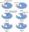
Schematic representation of different types of binding pockets. The protein kinase is shown in blue, with the Asp-Phe-Gly (DFG) motif in green. Red color denotes the aspartate amino acid residue of the DFG motif. The particular regions where different types of inhibitors bind are described below, the allosteric pocket is only a visualization and its place can be anywhere outside the ATP binding site.
Figure 2
Figure 2
Graphical illustration of different types of kinase inhibitors and their mode of action. Dark red hexagon represents an inhibitor. The protein kinase is shown in blue, the DFG motif in green, the aspartate amino acid residue of the DFG motif in red. In 2015 Wu demonstrated that co-crystal structure of 3-phosphoinositide-dependent protein kinase 1 (PDPK1, PDK1) with ATP showed that type I inhibitors interact with the active conformation of the enzyme where the aspartate residue of the DFG motif points into the ATP binding pocket, while type II inhibitors stabilize the inactive conformation of the enzyme where the aspartate residue faces outward of the binding site (PDB entry: 4RRV). Type III inhibitors interact with the allosteric site within the ATP binding pocket. Type IV inhibitors interact with the allosteric site. However, the allosteric pocket is only a visualization and its place can be anywhere outside the ATP binding site. Type V inhibitors interact with both the allosteric and ATP binding pockets. Type VI inhibitors form covalent bonds with either the ATP binding pocket or the allosteric pocket.
Figure 3
Figure 3
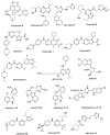
Chemical structures of some of the most studied type I cyclin-dependent kinase (CDK) inhibitors.
Figure 4
Figure 4
Chemical structure of quinoline-based type 1.5 inhibitor of monomeric CDK2.
Figure 5
Figure 5
Chemical structures of some of the most studied type II CDK inhibitors.
Figure 6
Figure 6
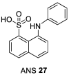
Chemical structure of 8-anilino-1-naphthalene sulfonate (ANS) the most studied type IV CDK inhibitor.
Figure 7
Figure 7
Chemical structures of some of the most studied type VI CDK inhibitors.
Figure 8
Figure 8
Chemical structure of CR8. A surface-exposed 2-pyridyl moiety of CR8 is responsible for glue degrader properties.
Figure 9
Figure 9
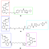
Chemical structures of CDK4/6 PROteolysis TArgeting Chimeras (PROTACs). Red rectangle denotes the palbociclib moiety, green rectangle denotes the ribociclib moiety, light blue rectangle denotes the thalidomide moiety and dark blue rectangle denotes the pomalidomide moiety.
Figure 10
Figure 10
Chemical structure of CDK8 PROTAC. Red rectangle denotes the pomalidomide moiety, light blue rectangle denotes the Cortistatin A derivative JH-VIII-49 moiety.
Figure 11
Figure 11
Chemical structures of CDK9 PROTACs. Red rectangle denotes the Thalidomide moiety, dark blue rectangle denotes the Pomalidomide moiety, green rectangle denotes the aminopyrazole derivative moiety, light blue rectangle denotes the SNS-032 moiety and violet rectangle denotes the Wogonin moiety.

Similar articles

Cited by

References

    1. Pavletich N.P. Mechanisms of cyclin-dependent kinase regulation: Structures of cdks, their cyclin activators, and cip and INK4 inhibitors. J. Mol. Biol. 1999;287:821–828. doi: 10.1006/jmbi.1999.2640. - DOI - PubMed
    1. Cohen P. Protein kinases—The major drug targets of the twenty-first century? Nat. Rev. Drug Discov. 2002;1:309–315. doi: 10.1038/nrd773. - DOI - PubMed
    1. Marra A., Curigliano G. Are all cyclin-dependent kinases 4/6 inhibitors created equal? NPJ Breast Cancer. 2019;5:27. doi: 10.1038/s41523-019-0121-y. - DOI - PMC - PubMed
    1. Wu P., Clausen M.H., Nielsen T.E. Allosteric small-molecule kinase inhibitors. Pharmacol. Ther. 2015;156:59–68. doi: 10.1016/j.pharmthera.2015.10.002. - DOI - PubMed
    1. Morgan D.O. The Cell Cycle: Principles of Control. 1st ed. New Science Press; London, UK: 2007.

MeSH terms

LinkOut - more resources