. 2021 Mar 10;11(3):190.
doi: 10.3390/jpm11030190.

Structured Integration and Alignment Algorithm: A Tool for Personalized Surgical Treatment of Tibial Plateau Fractures

Affiliations

Structured Integration and Alignment Algorithm: A Tool for Personalized Surgical Treatment of Tibial Plateau Fractures

Flaviu Moldovan et al. J Pers Med. .

Abstract

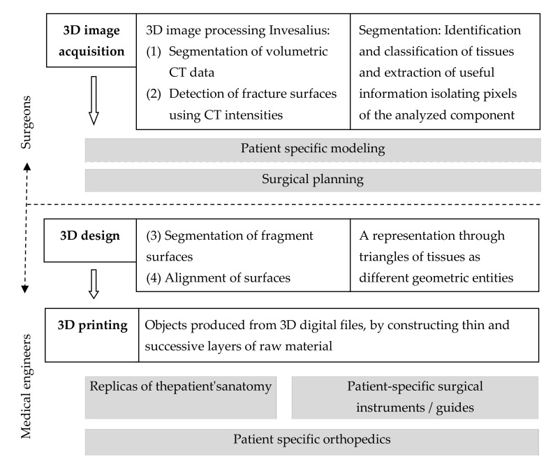
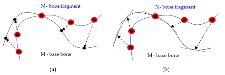
The planning of the surgical treatment in orthopedics, with the help of three-dimensional (3D) technologies, arouses an increasing scientific interest. Scientific literature describes some semi-automatic reconstructive attempts at fragmented bone fractures, but the matching algorithms presented are likely to improve. The aim of this paper is to develop a new method of aligning fragments of comminutive fractures. We have created a structured integration process and an alignment algorithm integrated in a clinical workflow for personalized surgical treatment of fractures. The provided solution is able to align the surfaces of bone fragments derived from the segmentation process of volumetric tomographic data. Positional uncertainties are eliminated interactively by the user, who selects the corresponding pairs of fracture surfaces. The final matching and the right alignment are performed automatically by the innovative alignment algorithm. The paper solves a challenging problem for the reconstruction of fractured bones, namely the choice of the optimal matching option from the situation in which surface portions of a fracture fragment correspond to multiple high fragments. The method is validated in practice for preoperative planning of a 49-year-old male patient who had a tibial plateau fracture of Schatzker type VI.

Keywords: alignment algorithm; orthopedic surgery; personalized treatment; three-dimensional printing; tibial fracture; workflow.


Conflict of interest statement

The authors declare no conflict of interest.

Figures

Figure 1
Figure 1
Clinical workflow for structured integration process supported by an alignment algorithm for personalized surgical treatment of fractures.
Figure 2
Figure 2
Comminutive tibial fractures shown in CT sections used in the process of the bone fragment fracture surfaces identification. The bone fragment segmented surface is delimited in color: (a) the inside of the bone has a higher intensity than that of the cortex; (b) the estimated surface segmentation is at a distance from the cortical surface.
Figure 3
Figure 3
Cortical and cancellous tissue view in a CT: (a) high-intensity circular regions show thick cortical bone tissue in the median area of the tibia; (b) more proximal, there is much less contrast between the regions of cortical and cancellous bone tissue.
Figure 4
Figure 4
(a) Point-to-point method: points are matched to discrete points; (b) Point-to-plane method: points are suitable for continuous tangent planes.
Figure 5
Figure 5
Matching the points between the N and M nodes of two surfaces (defined by the meshes of triangular networks). Vectors in red indicate matches that should be avoided, as they will cause the N surface to be erroneously shifted up and to the left.
Figure 6
Figure 6
The networks M and N are cut at the common boundary: (a) the intersection between the edges of the M and N meshes allows determination of triangle intersection points; (b) portions of triangles on the M and N meshes are removed; (c) both networks are limited at the points of intersection and new triangles are formed.
Figure 7
Figure 7
Extended network boundary for cutting in three-dimensional space.
Figure 8
Figure 8
(a) Digital Imaging and Communications in Medicine (DICOM) image; and (b) RadiAnt DICOM image of the patient included in the study.
Figure 9
Figure 9
Tibial plateau fracture Schatzker type VI: (a) posterior view of the 3D segmented model; (b) lateral view of the 3D segmented model; (c) posterior view of the 3D printed replica; (d) lateral view of the 3D printed replica.
Figure 10
Figure 10
Tibial plateau fracture Schatzker type VI—identification of independent fracture fragments by staining of distinct fracture fragments: (a) posterior view; (b) lateral view.
Figure 11
Figure 11
Tibial plateau fracture reconstruction result supported by the alignment algorithm: (a) posterior view of the standard triangle language (STL) model; (b) lateral view of the STL model; (c) posterior view of the 3D printed replica; (d) posterior view of the 3D printed replica.
Figure 12
Figure 12
Evaluation of the tibial plateau fracture reduction process by measuring the distances between the fracture fragments and the base bone: (a) initial fracture before reduction; (b) fracture after reduction supported by the alignment algorithm.
Figure 13
Figure 13
Variation of distances between bone fragments along the fracture line before (curve in red) and after alignment (curve in blue).
Figure 14
Figure 14
Assessment of the alignment method performance for three values of the evaluation criteria, the mean distance between fragments bones: (a) 0.5 mm, (b) 0.7 m and (c) 1 mm.

References

    1. Younes N., Syed N., Yadav S.K., Haris M., Abdallah A.M., Abu-Madi M. A Whole-Genome Sequencing Association Study of Low Bone Mineral Density Identifies New Susceptibility Loci in the Phase I Qatar Biobank Cohort. J. Pers. Med. 2021;11:34. doi: 10.3390/jpm11010034. - DOI - PMC - PubMed
    1. Mazurenko E., Rymar O., Shcherbakova L., Mazdorova E., Malyutina S. The Risk of Osteoporotic Forearm Fractures in Postmenopausal Women in a Siberian Population Sample. J. Pers. Med. 2020;10:77. doi: 10.3390/jpm10030077. - DOI - PMC - PubMed
    1. Curtin P., Conway A., Martin L., Lin E., Jayakumar P., Swart E. Compilation and Analysis of Web-Based Orthopedic Personalized Predictive Tools: A Scoping Review. J. Pers. Med. 2020;10:223. doi: 10.3390/jpm10040223. - DOI - PMC - PubMed
    1. Tanzi L., Vezzetti E., Moreno R., Moos S. X-Ray Bone Fracture Classification Using Deep Learning: A Baseline for Designing a Reliable Approach. Appl. Sci. 2020;10:1507. doi: 10.3390/app10041507. - DOI
    1. Tanzi L., Vezzetti E., Moreno R., Aprato A., Audisio A., Massè A. Hierarchical fracture classification of proximal femur X-ray images using a multistage Deep Learning approach. Eur. J. Radiol. 2020;133:109373. doi: 10.1016/j.ejrad.2020.109373. - DOI - PubMed

LinkOut - more resources