. 2021 Mar 10;13(3):449.
doi: 10.3390/v13030449.

HERV-K Gag RNA and Protein Levels Are Elevated in Malignant Regions of the Prostate in Males with Prostate Cancer

Affiliations

HERV-K Gag RNA and Protein Levels Are Elevated in Malignant Regions of the Prostate in Males with Prostate Cancer

Simin D Rezaei et al. Viruses. .

Abstract

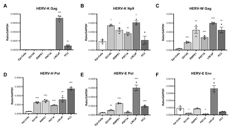
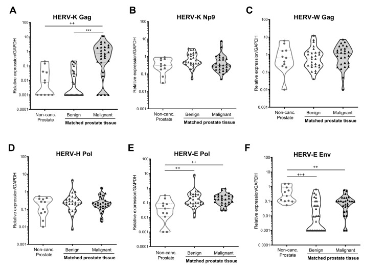
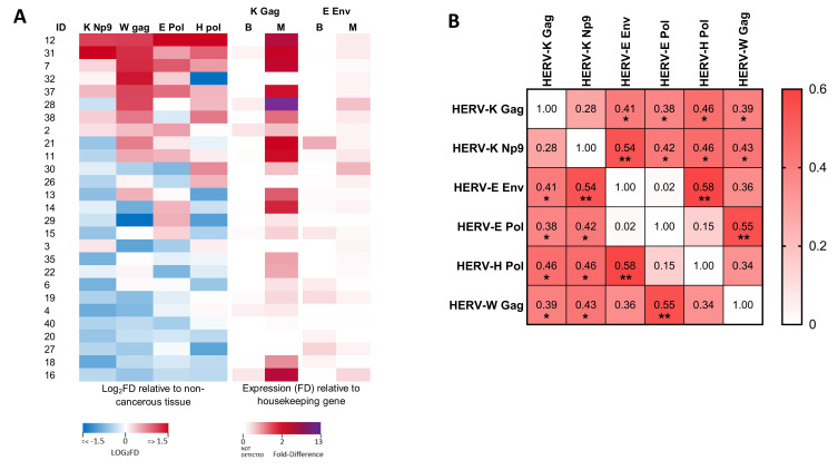
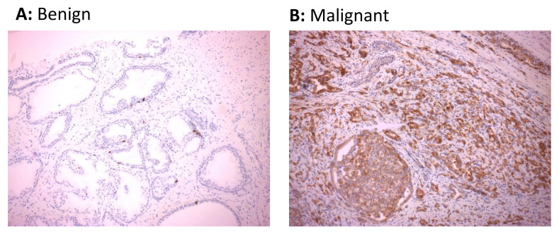
Heightened expression of human endogenous retrovirus (HERV) sequences has been associated with a range of malignancies, including prostate cancer, suggesting that they may serve as useful diagnostic or prognostic cancer biomarkers. We analysed the expression of HERV-K (Gag and Env/Np9 regions), HERV-E 4.1 (Pol and Env regions), HERV-H (Pol) and HERV-W (Gag) sequences in prostate cancer cells lines and normal prostate epithelial cells using qRT-PCR. HERV expression was also analysed in matched malignant and benign prostate tissue samples from men with prostate cancer (n = 27, median age 65.2 years (range 47-70)) and compared to prostate cancer-free male controls (n = 11). Prostate cancer epithelial cell lines exhibited a signature of HERV RNA overexpression, with all HERVs analysed, except HERV-E Pol, showing heightened expression in at least two, but more commonly all, cell lines analysed. Analysis of primary prostate material indicated increased expression of HERV-E Pol but decreased expression of HERV-E Env in both malignant and benign regions of the prostate in men with prostate cancer as compared to those without. Expression of HERV-K Gag was significantly higher in malignant regions of the prostate in men with prostate cancer as compared to matched benign regions and prostate cancer-free men (p < 0.001 for both), with 85.2% of prostate cancers donors showing malignancy-associated upregulation of HERV-K Gag RNA. HERV-K Gag protein was detected in 12/18 (66.7%) malignant tissues using immunohistochemistry, but only 1/18 (5.6%) benign tissue sections. Heightened expression of HERV-K Gag RNA and protein appears to be a sensitive and specific biomarker of prostate malignancy in this cohort of men with prostate carcinoma, supporting its potential utility as a non-invasive, adjunct clinical biomarker.

Keywords: HERV-K; human endogenous retrovirus (HERV); prostate cancer.


Conflict of interest statement

The authors declare no conflict of interest. The funders had no role in the design of the study; in the collection, analyses, or interpretation of data; in the writing of the manuscript, or in the decision to publish the results.

Figures

Figure 1
Figure 1
HERV transcript expression in prostate cancer cell lines and primary prostate epithelial cells. Expression of HERV-K Gag (A), HERV-K Np9 (B), HERV-W Gag (C), HERV-H Pol (D), HERV-E Pol (E) and HERV-E Env (F) transcripts were detected in cDNA synthesised from RNA purified from primary non-cancerous prostate epithelial cells (Epi-cells), DU145, RWPE1, PNT1A, LNCaP and PC3 cells by qPCR. Copies of HERV transcripts were standardised to the house-keeping gene GAPDH detected within the same samples. Graphs show the mean and standard error of the mean of results from n = 3 independent experiments. *, **, and *** indicate p < 0.05, 0.01, and 0.001 respectively, compared to primary epithelial cells, as determined by Student’s unpaired t-test.
Figure 2
Figure 2
HERV transcript expression in prostate biopsy samples from males with and without prostate cancer. Expression of HERV-K Gag (A), HERV-K Np9 (B), HERV-W Gag (C), HERV-H Pol (D), HERV-E Pol (E) and HERV-E Env (F) transcripts were detected in RNA extracted from punch biopsy prostate samples from men without prostate cancer (Non-Canc. Prostate, n = 11), or from matched benign and malignant regions of the prostate from individuals with prostate cancer (n = 27) by qRT-PCR. Graphs show truncated violin plots of the average gene expression standardised to copies of GAPDH from n = 3 replicates per sample. Samples with undetectable levels of HERV transcript are shown with a value of 0.001 for the purposes of visualisation in the graphs, and the actual value of 0 was used for statistical analyses. ++, and +++ indicate p < 0.01 and 0.001 respectively, as compared to non-cancerous prostate tissue, determined by Mann–Whitney U-test. *** indicates p < 0.001 for comparison of matched benign and malignant tissue, as determined by Wilcoxon matched-pairs signed-rank test. Non-parametric tests were used due to non-normal distribution of the data.
Figure 3
Figure 3
Associations between expression of various HERV transcripts in prostate cancer tissue. (A) Heat map showing the differential expression (log2 fold difference; FD) of HERV RNA transcripts in prostate cancer tissues from men with prostate cancer. Left panels show expression of HERV-K Np9, HERV-W Gag, HERV-E Pol and HERV-H Pol in malignant regions relative to benign regions of the prostate (each standardised to GAPDH). Right panels show expression of HERV-K Gag and HERV-E Env in both benign (B) and malignant (M) regions relative to expression of GAPDH (due to transcripts being undetectable in many benign tissue samples). (B) Correlation matrix showing the association between expression levels of the various HERV transcripts as determined by non-parametric Spearman’s correlation. For HERV-K Gag and HERV-E Env, the expression level in malignant tissues (relative to GAPDH) was analysed, whilst for all other transcripts, the expression in malignant tissue relative to benign tissue (both standardised to GAPDH) was used. Values show Spearman’s rho. * and ** indicate p < 0.05 and 0.01, respectively.
Figure 4
Figure 4
Expression of HERV-K Gag protein in cancerous regions of prostate tissue sections. HERV-K Gag protein was detected in sections of matched malignant and benign regions of formalin-fixed paraffin-embedded prostate biopsy tissue from 18 males with prostate cancer by immunohistochemistry. Immunohistochemical images from benign prostate tissue (A) or malignant prostatic adenocarcinoma (B) regions of the prostate showing negative and positive staining for HERV-K Gag protein from one representative individual are shown.

Similar articles

Cited by

References

    1. Australian Institute of Health and Welfare Cancer Data in Australia. [(accessed on 12 February 2021)]; Available online: https://www.aihw.gov.au/reports/cancer/cancer-data-in-australia/contents....
    1. Prostate Cancer Foundation of Australia Prostate Cancer in Australia—What Do the Numbers Tell Us? [(accessed on 12 February 2021)]; Available online: https://www.prostate.org.au/news-media/news/prostate-cancer-in-australia...
    1. Glasziou P.P., Jones M.A., Pathirana T., Barratt A.L., Bell K.J. Estimating the magnitude of cancer overdiagnosis in Australia. Med. J. Aust. 2020;212:163–168. doi: 10.5694/mja2.50455. - DOI - PMC - PubMed
    1. Geis F.K., Goff S.P. Silencing and transcriptional regulation of endogenous retroviruses: An overview. Viruses. 2020;12:884. doi: 10.3390/v12080884. - DOI - PMC - PubMed
    1. Buscher K., Hahn S., Hofmann M., Trefzer U., Ozel M., Sterry W., Lower J., Lower R., Kurth R., Denner J. Expression of the human endogenous retrovirus-K transmembrane envelope, Rec and Np9 proteins in melanomas and melanoma cell lines. Melanoma Res. 2006;16:223–234. doi: 10.1097/01.cmr.0000215031.07941.ca. - DOI - PubMed

Publication types

MeSH terms

LinkOut - more resources