. 2021 Mar 10;11(3):410.
doi: 10.3390/biom11030410.

Actin-Resistant DNase1L2 as a Potential Therapeutics for CF Lung Disease

Affiliations

Actin-Resistant DNase1L2 as a Potential Therapeutics for CF Lung Disease

Danila Delfino et al. Biomolecules. .

Abstract

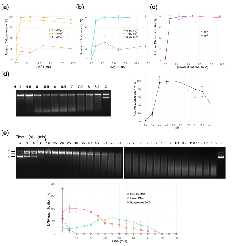
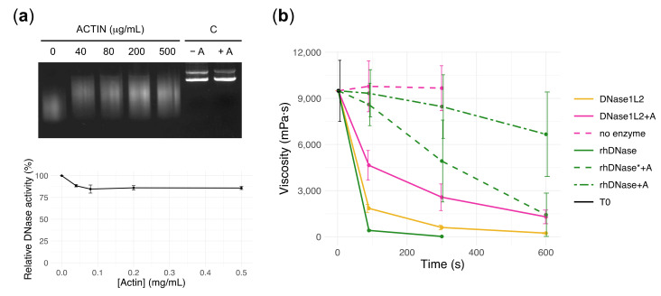
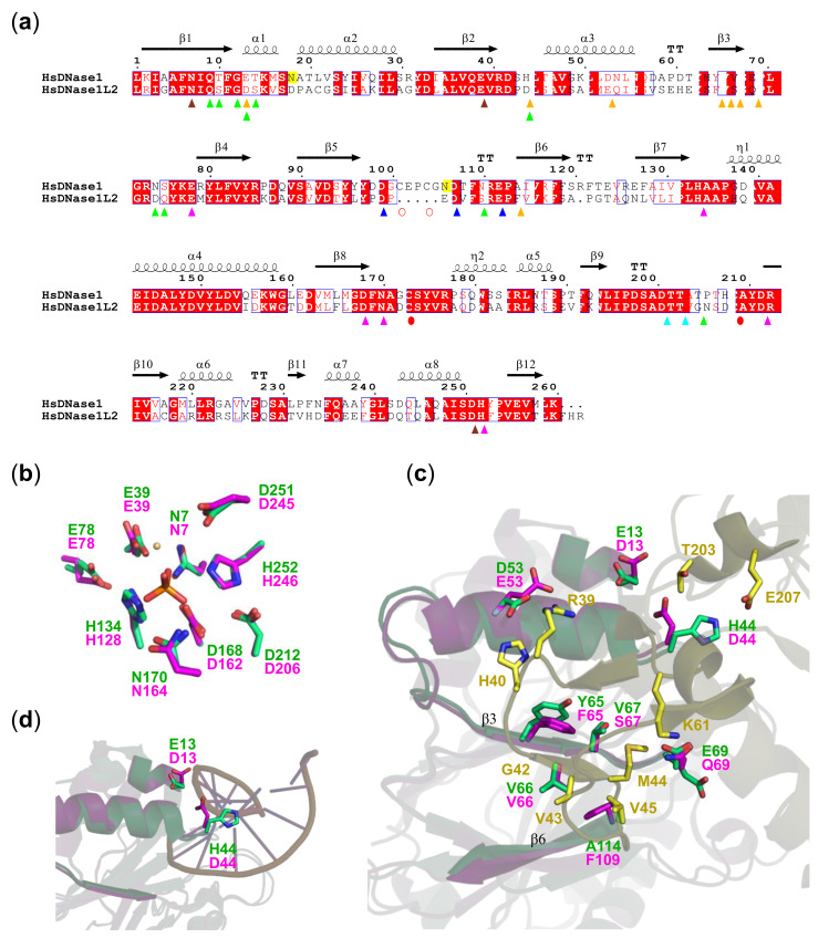
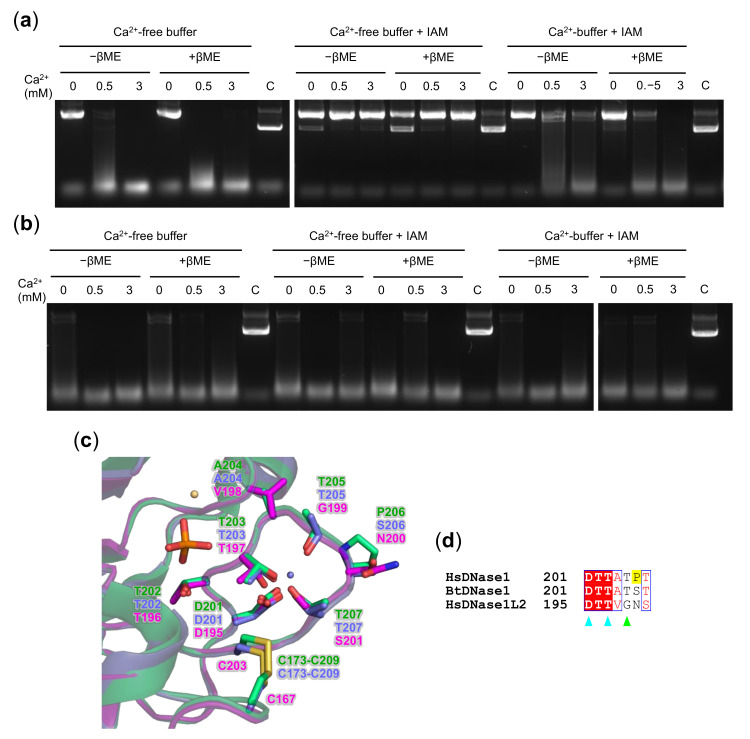
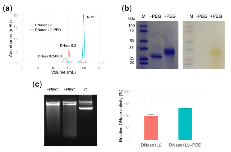
In cystic fibrosis (CF), the accumulation of viscous lung secretions rich in DNA and actin is a major cause of chronic inflammation and recurrent infections leading to airway obstruction. Mucolytic therapy based on recombinant human DNase1 reduces CF mucus viscosity and promotes airway clearance. However, the marked susceptibility to actin inhibition of this enzyme prompts the research of alternative treatments that could overcome this limitation. Within the human DNase repertoire, DNase1L2 is ideally suited for this purpose because it exhibits metal-dependent endonuclease activity on plasmid DNA in a broad range of pH with acidic optimum and is minimally inhibited by actin. When tested on CF artificial mucus enriched with actin, submicromolar concentrations of DNase1L2 reduces mucus viscosity by 50% in a few seconds. Inspection of superimposed model structures of DNase1 and DNase1L2 highlights differences at the actin-binding interface that justify the increased resistance of DNase1L2 toward actin inhibition. Furthermore, a PEGylated form of the enzyme with preserved enzymatic activity was obtained, showing interesting results in terms of activity. This work represents an effort toward the exploitation of natural DNase variants as promising alternatives to DNase1 for the treatment of CF lung disease.

Keywords: PEGylation; Pichia pastoris; cystic fibrosis; endonuclease; enzyme therapeutics; mucolytics.


Conflict of interest statement

The authors declare no conflict of interest. The funders had no role in the design of the study; in the collection, analyses, or interpretation of data; in the writing of the manuscript, or in the decision to publish the results.

Figures

Figure 1
Figure 1
Expression and purification of DNase1L2. (a) Portion of multiple alignment showing the proline-rich region present only in some mammalian DNase1L2 sequences (names in blue). Vertical lines on the left indicate sequence from mammals (pink) and sauropsids (brown). (b) Frequency plot of the aligned proline-rich regions from thirteen mammalian DNase1L2 sequences showing remarkable conservation. (c) Scheme of the DNase1L2 mRNA sequence (NM_001374.3) and the constructs designed for the expression of DNase1L2-L and DNase1L2-S in P. pastoris GS115. CDS lacking the predicted signal peptide were cloned into pPIC9K vector bearing a sequence (α-factor) for the secretion of expressed proteins. (d) SDS-PAGE analysis of extracellular expression of P. pastoris GS115 cells transformed with pPIC9K-DNase1L2-L (two clones, 1 and 2, are shown) or with pPIC9K-DNase1L2-S (two clones, 3 and 4, are shown) or with an empty vector (EV). The band corresponding to DNase1L2-S is found at the expected molecular weight of 28.8 kDa. M: Marker. (e) Size-exclusion chromatogram and SDS-PAGE (inset) showing the final step of DNase1L2 purification. Numerals above peaks correspond to the gel lanes in the inset. L: Load.
Figure 2
Figure 2
Characterization of DNase1L2 activity using purified plasmid DNA. (ac) Gel densitometric analysis showing the effects of divalent cations on DNase1L2 activity. Data are means ± SD of three independent experiments. (a) Calcium titration in the presence of 0, 1, and 3 mM MgCl2. (b) Magnesium titration in the presence of 0, 1, and 3 mM CaCl2. (c) Manganese titration and cobalt titration. (d) Agarose gel electrophoresis (left panel) and densitometric analysis (right panel) showing the effect of pH on DNase1L2 activity. C: control, plasmid DNA without enzyme at pH 4. Data are means ± SD of three independent experiments. (e) Agarose gel electrophoresis (upper panel) and densitometric analysis (bottom panel) showing the time course of DNase1L2 activity. Plasmid DNA (200 ng; 90% supercoiled) was incubated with DNase1L2 (69 pmol) in the time range from 1 s to 125 min. C: control, supercoiled plasmid DNA without enzyme. Plasmid DNA forms are indicated at the left of the gel and correspond to the plots in the graph; C: circular DNA, L: linear DNA, S: supercoiled DNA. Data are means ± SD of two independent experiments.
Figure 3
Figure 3
DNase1L2 reduces viscosity of cystic fibrosis (CF) artificial mucus with marked resistance to actin inhibition. (a) Agarose gel electrophoresis (upper panel) and densitometric analysis (bottom panel) showing the effect of actin on plasmid DNA digestion by DNase1L2. C: control, purified plasmid DNA without enzyme in the absence (-A) and in the presence of actin (+A). Data are means ± SD of two independent experiments. (b) Viscosity measurements showing the activity of DNase1L2 and rhDNase in CF artificial mucus (CF-AM). Activity of DNase1L2 (5 units: 20 ng/μL, pink solid and dashed line) and rhDNase (5 units: 2 ng/μL, green solid and dashed line, or 50 U: 20 ng/μL, green dashed-dot line) was tested in the presence (0.2 mg/mL) or absence of actin (A). T0 is CF-AM at the beginning of measurement. Data are means ± SD of at least two independent experiments. Viscosity values (mPa·s; mean ± SD) are reported in Table S2.
Figure 4
Figure 4
DNase1L2 exhibits conservation of DNase1 active site and diversity of the actin-binding region. (a) Aligned sequences of human DNase1 (HsDNase1) and DNase1L2 (HsDNase1L2). The residue numbering refers to the DNase1 sequence and is the same as in the structure in (bd). The signal sequence that is cleaved prior to secretion in both DNases is not shown. Secondary structure elements inferred from 4AWN are shown above the alignment. Remarkable residues are indicated as follows: catalytic, pink triangles; Mg2+ binding, brown triangles; Ca2+ binding in site I, cyan triangles; Ca2+ binding in site II, blue triangles; actin binding, orange triangles; DNA binding, green triangles; conserved cysteines, red circles; cysteines present only in DNase1, open red circles; and N-linked glycosylation sites are highlighted in yellow. (b) Superimposition of the active sites of DNase1L2 homology model (pink carbons) and human DNase1 structure (4AWN; green carbons). Bound Mg2+ (light orange) and phosphate (orange) ions are shown. (c) Superimposition of the DNase1L2 homology model (pink carbons) with the human DNase1 structure (4AWN; green carbons) and the actin-bovine DNase1 complex (1ATN; yellow carbons), showing the actin-binding region. The key residues involved in interactions are shown in sticks with labels. β-strands of DNase1L2 and DNase1 interacting with actin are labeled. (d) Superimposition of the DNase1L2 homology model (pink carbons) with 4AWN (green carbons) and the d(GGTATACC)2-bovine DNase1 complex (1DNK; magenta carbons), showing the DNA-binding region. The key residues involved in interactions are shown in sticks with labels.
Figure 5
Figure 5
Ca2+ protection of DNase1L2 reduced cysteines from alkylation. Agarose gel electrophoresis showing the effect of β–mercaptoethanol (βME) and iodoacetamide (IAM) on plasmid DNA digestion by (a) DNase1L2 and (b) rhDNase. The enzymes were in buffer supplemented with 1 mM CaCl2 or in CaCl2-free buffer. After treatment, reactions were assembled as described in Materials and Methods. (c) Superimposition of the DNase1L2 model (pink carbons) with the human DNase1 structure (4AWN; green carbons) and the bovine DNase1 structure (1ATN; violet carbons), showing the calcium-binding site I. Bound Ca2+ (violet), Mg2+ (light orange), and phosphate (orange) ions are shown. (d) Aligned sequences of human DNase1 (HsDNase1), bovine DNase1 (BtDNase1), and human DNase1L2 (HsDNase1L2). The position of the first amino acid of each sequence is on the right. The residue numbering is the same as in the structure in (c). Key residues are indicated as follows: Ca2+-binding site I, cyan triangles; DNA binding, green triangle; and the proline (P206) discussed in the text is highlighted in yellow.
Figure 6
Figure 6
PEGylation of DNase1L2. (a) Size-exclusion chromatogram of DNase1L2 (salmon) and reaction mixture of DNase1L2 and PEG-NHS (cyan). Representative peaks are labelled. (b) SDS-PAGE of native and PEGylated DNase1L2 with Coomassie (left panel) and PEG-specific barium iodide dye (right panel) staining. M: marker. (c) Agarose gel electrophoresis (left panel) and densitometric analysis (right panel) showing DNase1L2 and DNase1L2-PEG activity on supercoiled plasmid DNA. Data are means ± SD of two independent experiments.

Similar articles

Cited by

References

    1. Elborn J.S. Cystic fibrosis. Lancet. 2016;388:2519–2531. doi: 10.1016/S0140-6736(16)00576-6. - DOI - PubMed
    1. Mall M.A., Hartl D. CFTR: Cystic fibrosis and beyond. Eur. Respir. J. 2014;44:1042–1054. doi: 10.1183/09031936.00228013. - DOI - PubMed
    1. Davis P.B., Drumm M., Konstan M.W. Cystic fibrosis. Am. J. Respir. Crit. Care Med. 1996;154:1229–1256. doi: 10.1164/ajrccm.154.5.8912731. - DOI - PubMed
    1. Davis P.B. Cystic fibrosis since 1938. Am. J. Respir. Crit. Care Med. 2006;173:475–482. doi: 10.1164/rccm.200505-840OE. - DOI - PubMed
    1. Lethem M.I., James S.L., Marriott C., Burke J.F. The origin of DNA associated with mucus glycoproteins in cystic fibrosis sputum. Eur. Respir. J. 1990;3:19–23. - PubMed

Publication types

MeSH terms

LinkOut - more resources