Review
. 2021 Mar 10;11(3):225.
doi: 10.3390/life11030225.

Recent Progress Using De Novo Design to Study Protein Structure, Design and Binding Interactions

Affiliations
Review

Recent Progress Using De Novo Design to Study Protein Structure, Design and Binding Interactions

Juan Ferrando et al. Life (Basel). .

Abstract

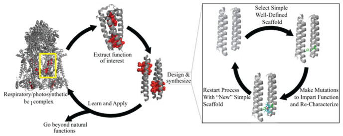
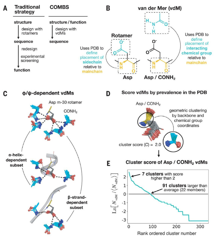
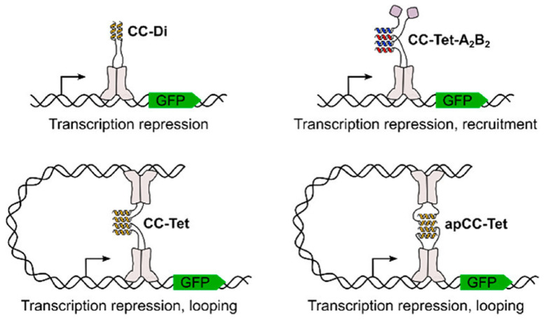
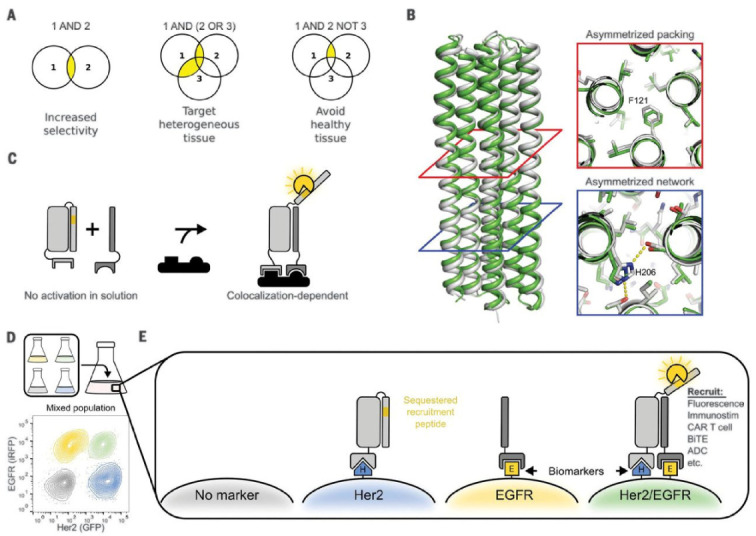
De novo protein design is a powerful methodology used to study natural functions in an artificial-protein context. Since its inception, it has been used to reproduce a plethora of reactions and uncover biophysical principles that are often difficult to extract from direct studies of natural proteins. Natural proteins are capable of assuming a variety of different structures and subsequently binding ligands at impressively high levels of both specificity and affinity. Here, we will review recent examples of de novo design studies on binding reactions for small molecules, nucleic acids, and the formation of protein-protein interactions. We will then discuss some new structural advances in the field. Finally, we will discuss some advancements in computational modeling and design approaches and provide an overview of some modern algorithmic tools being used to design these proteins.

Keywords: binding; de novo protein design; protein-protein interactions.


Conflict of interest statement

The authors declare no conflict of interest.

Figures

Figure 1
Figure 1
Process flow of De Novo protein design. Starting with a specific protein, the function of interest is isolated. A protein is modified from basic chemical principles so that it reproduces that function. In so doing, the designers can learn and rigorously test the underlying biophysical principles. The design and synthesis process itself begins with a simple scaffold. Mutations are made to impart function and the whole protein is recharacterized. If function is not achieved, or achieved to a sufficient level, the process restarts, however the modified protein is the new scaffold. In this iterative process complexity is kept to a minimum. An important aspect of de novo design is that proteins can be used in abiological contexts. This allows the expansion of natural functions into areas of synthetic chemistry making the de novo proteins a versatile tool capable of addressing many issues.
Figure 2
Figure 2
A depiction of the workflow of the new van der Mer unit. (A) The classical workflow of traditional protein design versus the COMBS methodology described in the paper [40]. (B) Definition of the van der Mer unit, accounting for the distance between the backbone Cα and the small molecule chemical group. (C) Next step of van der Mer modeling highlighting the rotamer and ϕ and ψ angle dependence. (D,E) Ranking of the prevalence of the chemical group-protein pair in the PDB and cluster score. The ideal amino acid side chain based on this analysis, and considering other possible interactions in the scaffold, is then selected for analysis. This work was reprinted with permission from: A defined structural unit enables de novo design of small-molecule-binding proteins. Polizzi, N.F., DeGrado, W.F. Science 2020, 369, 1227–1233. Copyright (2020) AAAS.
Figure 3
Figure 3
Using protein-protein interactions to rescue the function of a DNA binding protein. Edgell and coauthors attached their associating helices to the segments of the LacI repressor. Depending on the sequence of the de novo helical pairs the authors could form either two or four helix bundles, which provided them a method of tuning the function of their engineered repressor protein. Reprinted with permission from C. Edgell, L., Smith, A.J., Beesley, J.L., Savery, N.J. and Woolfson, D.N. De Novo Designed Protein-Interaction Modules for In-Cell Applications. ACS Synth Biol. Volume 9, no. 2, pp. 427–436, February 2020.
Figure 4
Figure 4
Incorporation of protein logic with the LOCKR system of proteins. (A) Combinations of protein-protien interactions that lead to different logic functions. (B) Structure of the scaffold “cage” protein used to create the Co-LOCKR system. (C) Colocalization of proteins is required for detection. Proteins are designed to not interact significantly in solution, but only when they are colocalized. (D) Flow cytometry can discriminate cells based on their surface antigens due to the Co-LOCKR system. (E) Depiction of effector protein recruitment based on surface antigens. Figure subunits F and G removed for clarity. Reprinted with permission from Lajoie, M.J. et al. Designed protein logic to target cells with precise combinations of surface antigens. Science Volume 369, no. 6511, pp. 1637–1643, September 2020.

Similar articles

Cited by

References

    1. Vuignier K., Schappler J., Veuthey J.-L., Carrupt P.-A., Martel S. Drug–protein binding: A critical review of analytical tools. Anal. Bioanal. Chem. 2010;398:53–66. doi: 10.1007/s00216-010-3737-1. - DOI - PubMed
    1. Siggers T., Gordân R. Protein–DNA binding: Complexities and multi-protein codes. Nucleic Acids Res. 2014;42:2099–2111. doi: 10.1093/nar/gkt1112. - DOI - PMC - PubMed
    1. Feng Y., Walsh C.A. Protein–Protein interactions, cytoskeletal regulation and neuronal migration. Nat. Rev. Neurosci. 2001;2:408–416. doi: 10.1038/35077559. - DOI - PubMed
    1. Dutton P.L., Moser C.C. Engineering enzymes. Faraday Discuss. 2011;148:443–448. doi: 10.1039/C005523A. - DOI - PMC - PubMed
    1. Lichtenstein B.R., Farid T.A., Kodali G., Solomon L.A., Anderson J.R., Sheehan M.M., Ennist N.M., Fry B.A., Chobot S.E., Bialas C., et al. Engineering oxidoreductases: Maquette proteins designed from scratch. Biochem. Soc. Trans. 2012;40:561–566. doi: 10.1042/BST20120067. - DOI - PMC - PubMed

LinkOut - more resources