Cache Domain Containing 1 Is a Novel Marker of Non-Alcoholic Steatohepatitis-Associated Hepatocarcinogenesis
- PMID: 33802238
- PMCID: PMC8001421
- DOI: 10.3390/cancers13061216
Cache Domain Containing 1 Is a Novel Marker of Non-Alcoholic Steatohepatitis-Associated Hepatocarcinogenesis
Abstract
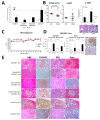
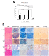
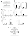
In the present study, potential molecular biomarkers of NASH hepatocarcinogenesis were investigated using the STAM mice NASH model, characterized by impaired insulin secretion and development of insulin resistance. In this model, 2-days-old C57BL/6N mice were subjected to a single subcutaneous (s.c.) injection of 200 μg streptozotocin (STZ) to induce diabetes mellitus (DM). Four weeks later, mice were administered high-fat diet (HFD) HFD-60 for 14 weeks (STAM group), or fed control diet (STZ group). Eighteen-week-old mice were euthanized to allow macroscopic, microscopic, histopathological, immunohistochemical and proteome analyses. The administration of HFD to STZ-treated mice induced significant fat accumulation and fibrosis development in the liver, which progressed to NASH, and rise of hepatocellular adenomas (HCAs) and carcinomas (HCCs). In 18-week-old animals, a significant increase in the incidence and multiplicity of HCAs and HCCs was found. On the basis of results of proteome analysis of STAM mice HCCs, a novel highly elevated protein in HCCs, cache domain-containing 1 (CACHD1), was chosen as a potential NASH-HCC biomarker candidate. Immunohistochemical assessment demonstrated that STAM mice liver basophilic, eosinophilic and mixed-type altered foci, HCAs and HCCs were strongly positive for CACHD1. The number and area of CACHD1-positive foci, and cell proliferation index in the area of foci in mice of the STAM group were significantly increased compared to that of STZ group. In vitro siRNA knockdown of CACHD1 in human Huh7 and HepG2 liver cancer cell lines resulted in significant inhibition of cell survival and proliferation. Analysis of the proteome of knockdown cells indicated that apoptosis and autophagy processes could be activated. From these results, CACHD1 is an early NASH-associated biomarker of liver preneoplastic and neoplastic lesions, and a potential target protein in DM/NASH-associated hepatocarcinogenesis.
Keywords: CACHD1; HCC; NASH; STAM mice; hepatocarcinogenesis.
Conflict of interest statement
The authors declare no conflict of interest.
Figures


Similar articles
-
Accumulation of 8-hydroxydeoxyguanosine, L-arginine and Glucose Metabolites by Liver Tumor Cells Are the Important Characteristic Features of Metabolic Syndrome and Non-Alcoholic Steatohepatitis-Associated Hepatocarcinogenesis.Int J Mol Sci. 2020 Oct 20;21(20):7746. doi: 10.3390/ijms21207746. Int J Mol Sci. 2020. PMID: 33092030 Free PMC article.
-
Hepatic expression of the Sptlc3 subunit of serine palmitoyltransferase is associated with the development of hepatocellular carcinoma in a mouse model of nonalcoholic steatohepatitis.Oncol Rep. 2015 Apr;33(4):1657-66. doi: 10.3892/or.2015.3745. Epub 2015 Jan 21. Oncol Rep. 2015. PMID: 25607821
-
Analysis of amino acid profiles of blood over time and biomarkers associated with non-alcoholic steatohepatitis in STAM mice.Exp Anim. 2019 Nov 6;68(4):417-428. doi: 10.1538/expanim.18-0152. Epub 2019 Jun 1. Exp Anim. 2019. PMID: 31155606 Free PMC article.
-
Proteome Characteristics of Non-Alcoholic Steatohepatitis Liver Tissue and Associated Hepatocellular Carcinomas.Int J Mol Sci. 2017 Feb 17;18(2):434. doi: 10.3390/ijms18020434. Int J Mol Sci. 2017. PMID: 28218651 Free PMC article.
-
Pleiotropic effects of methionine adenosyltransferases deregulation as determinants of liver cancer progression and prognosis.J Hepatol. 2013 Oct;59(4):830-41. doi: 10.1016/j.jhep.2013.04.031. Epub 2013 May 7. J Hepatol. 2013. PMID: 23665184 Review.
Cited by
-
Recent Insights into the Biomarkers, Molecular Targets and Mechanisms of Non-Alcoholic Steatohepatitis-Driven Hepatocarcinogenesis.Cancers (Basel). 2023 Sep 14;15(18):4566. doi: 10.3390/cancers15184566. Cancers (Basel). 2023. PMID: 37760534 Free PMC article. Review.
-
Biallelic loss-of-function variants in CACHD1 cause a novel neurodevelopmental syndrome with facial dysmorphism and multisystem congenital abnormalities.Genet Med. 2024 Apr;26(4):101057. doi: 10.1016/j.gim.2023.101057. Epub 2023 Dec 27. Genet Med. 2024. PMID: 38158856 Free PMC article.
-
Unraveling the Janus-Faced Role of Autophagy in Hepatocellular Carcinoma: Implications for Therapeutic Interventions.Int J Mol Sci. 2023 Nov 13;24(22):16255. doi: 10.3390/ijms242216255. Int J Mol Sci. 2023. PMID: 38003445 Free PMC article. Review.
References
-
- Nakagawa H., Umemura A., Taniguchi K., Font-Burgada J., Dhar D., Ogata H., Zhong Z., Valasek M.A., Seki E., Hidalgo J., et al. ER stress cooperates with hypernutrition to trigger TNF-dependent spontaneous HCC development. Cancer Cell. 2014;26:331–343. doi: 10.1016/j.ccr.2014.07.001. - DOI - PMC - PubMed
Grants and funding
- 19710167/Ministry of Education, Culture, Sports and Science and Technology of Japan, Grant-in-Aid for Scientific Research
- 24501354/Ministry of Education, Culture, Sports and Science and Technology of Japan, Grant-in-Aid for Scientific Research
- Grant in Aid/Grant-in-Aid for Scientific Research from the Ministry of Health, Labour and Welfare of Japan
LinkOut - more resources
Full Text Sources
Other Literature Sources

