Case Reports
. 2021 Mar 3;18(5):2465.
doi: 10.3390/ijerph18052465.

Non-Incised Papilla Surgical Approach and Leukocyte Platelet-Rich Fibrin in Periodontal Reconstruction of Deep Intrabony Defects: A Case Series

Affiliations
Case Reports

Non-Incised Papilla Surgical Approach and Leukocyte Platelet-Rich Fibrin in Periodontal Reconstruction of Deep Intrabony Defects: A Case Series

Guillermo Pardo-Zamora et al. Int J Environ Res Public Health. .

Abstract

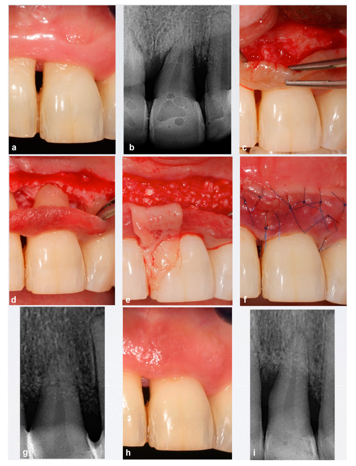
We present the preliminary results of the treatment of teeth with a deep, non-contained periodontal residual defect, vestibular bone dehiscence, and soft tissue recession, by combining an apical non-incised papilla surgical approach (NIPSA) to the defect and leukocyte platelet-rich fibrin (L-PRF) in the vestibular aspect. Four patients (upper left first premolar, upper left central incisor, upper right central incisor and upper right lateral incisor) have been treated. At one year of follow up, all cases showed a considerable reduction in the periodontal pocket depth, a gain in clinical attachment and no bleeding on probing, as well as an improvement in the marginal soft tissue minimizing soft tissue contraction (recession and/or loss of papilla) and improving soft tissue architecture. NIPSA plus L-PRF seem to improve clinical outcomes in deep non-contained intrabony defects associated with soft tissue recession.

Keywords: L-PRF; non-incised papilla surgical approach; periodontitis; reconstructive surgery; regeneration; surgical flaps.


Conflict of interest statement

The authors declare no conflict of interest.

Figures

Figure 1
Figure 1
Case 24. (a,b) Upper left first premolar with advanced periodontal destruction and grade II mobility with two mesial and distal intra-bone defects. (c) Apical incision exposing intrabony defects and lack of vestibular cortical plates. (d,e) Biomaterials and adapted platelet and leukocyte-rich fibrin (L-PRF) membrane. (f,g) Suture and post-surgical X-ray. (h) Closure at the first attempt seven days post-surgery. (ik) 12 months after surgery. A composite restoration was performed for aesthetic reasons. Significant reduction in probing pocket depth (PPD) and healthy soft tissues with acceptable scalloping. X-ray: resolution of intrabony defects.
Figure 2
Figure 2
Case 21. (a,b) Upper left central incisor with advanced periodontal destruction and grade II mobility with two mesial and distal intra-bone defects. (c,d) Apical incision exposing intrabony defects and lack of vestibular cortical plate. (e) Biomaterials and adapted L-PRF membrane. (f,g) Suture and post-surgical X-ray. (h,i) Clinical view and X-ray 12 months after surgery.
Figure 3
Figure 3
Case 11. (a,b) Upper right central incisor extruded with advanced periodontal destruction and grade II mobility with mesial intra-bone defect. (c,d) Suture and post-surgical X-ray. (e,f) 12 months after surgery. A composite restoration was performed for aesthetic reasons. X-ray: resolution of intrabony defect.
Figure 4
Figure 4
Case 12. (ac) Clinical and radiographic examinations revealed an upper right lateral incisor with distal intra-bone defect reaching the apex of the tooth. The soft tissue showed a nonscalloped architecture as a result of chronic inflammation. (d) Post-surgical X-ray. (e,f) 12 months after surgery. Harmonious scalloped gingiva with physiologically healthy interdental papillae height resulting in form-controlled inflammation and coronal displacement of the papillae. X-ray: resolution of intrabony defect.

Similar articles

Cited by

References

    1. Sculean A., Kiss A., Miliauskaite A., Schwarz F., Arweiler N.B., Hannig M. Ten-year results following treatment of intra-bony defects with enamel matrix proteins and guided tissue regeneration. J. Clin. Periodontol. 2008;35:817–824. doi: 10.1111/j.1600-051X.2008.01295.x. - DOI - PubMed
    1. Pretzl B., Kim T.-S., Steinbrenner H., Dörfer C., Himmer K., Eickholz P. Guided tissue regeneration with bioabsorbable barriers III 10-year results in infrabony defects. J. Clin. Periodontol. 2009;36:349–356. doi: 10.1111/j.1600-051X.2009.01378.x. - DOI - PubMed
    1. Prato G.P., Cortellini P. Thirty-year stability after regeneration of a deep intrabony defect: A case report. J. Clin. Periodontol. 2016;43:857–862. doi: 10.1111/jcpe.12587. - DOI - PubMed
    1. Wachtel H., Schenk G., Böhm S., Weng D., Zuhr O., Hürzeler M.B. Microsurgical access flap and enamel matrix derivative for the treatment of periodontal intrabony defects: A controlled clinical study. J. Clin. Periodontol. 2003;30:496–504. doi: 10.1034/j.1600-051X.2003.00013.x. - DOI - PubMed
    1. Cortellini P., Tonetti M.S. A minimally invasive surgical technique with an enamel matrix derivative in the regenerative treatment of intra-bony defects: A novel approach to limit morbidity. J. Clin. Periodontol. 2007;34:87–93. doi: 10.1111/j.1600-051X.2006.01020.x. - DOI - PubMed

Publication types

LinkOut - more resources