. 2021 Mar 3;9(3):517.
doi: 10.3390/microorganisms9030517.

Bacteriophages to Control Multi-Drug Resistant Enterococcus faecalis Infection of Dental Root Canals

Affiliations

Bacteriophages to Control Multi-Drug Resistant Enterococcus faecalis Infection of Dental Root Canals

Mohamed El-Telbany et al. Microorganisms. .

Abstract

Phage therapy is an alternative treatment to antibiotics that can overcome multi-drug resistant bacteria. In this study, we aimed to isolate and characterize lytic bacteriophages targeted against Enterococcus faecalis isolated from root canal infections obtained from clinics at the Faculty of Dentistry, Ismalia, Egypt. Bacteriophage, vB_ZEFP, was isolated from concentrated wastewater collected from hospital sewage. Morphological and genomic analysis revealed that the phage belongs to the Podoviridae family with a linear double-stranded DNA genome, consisting of 18,454, with a G + C content of 32.8%. Host range analysis revealed the phage could infect 10 of 13 E. faecalis isolates exhibiting a range of antibiotic resistances recovered from infected root canals with efficiency of plating values above 0.5. One-step growth curves of this phage showed that it has a burst size of 110 PFU per infected cell, with a latent period of 10 min. The lytic activity of this phage against E. faecalis biofilms showed that the phage was able to control the growth of E. faecalis in vitro. Phage vB_ZEFP could also prevent ex-vivo E. faecalis root canal infection. These results suggest that phage vB_ZEFP has potential for application in phage therapy and specifically in the prevention of infection after root canal treatment.

Keywords: Enterococcus faecalis; bacteriophage; multi-drug resistant; phage therapy; root canal infection.


Conflict of interest statement

The authors declare no conflict of interest. The funders had no role in the design of the study; in the collection, analyses, or interpretation of data; in the writing of the manuscript; or in the decision to publish the results.

Figures

Figure 1
Figure 1
Age distribution of oral carriers of E. faecalis.
Figure 2
Figure 2
Transmission electron micrograph of phage vB_ZEFP: The phage exhibits a short non-contractile tail indicative of the Podoviridae family. The scale bar represents 50 nm.
Figure 3
Figure 3
vB_ZEFP phage mediated reduction of E. faecalis biofilms. Microtitre plates containing preformed biofilms were treated either with phage for 24 h. Biofilm contents were estimated by OD measurement of ethanol solubilized crystal violet-stained material and reported as residual biofilm. MOI indicates the multiplicity of infection of the phage with respect to the initial E. faecalis viable count. All phage treatments reduced biofilm contents compared to the carrier treated controls (p < 0.003).
Figure 4
Figure 4
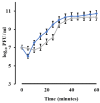
Single step growth curve. The grey line represents nascent phage without chloroform addition (PFU/mL), while the blue line represents phages released after chloroform addition (PFU/mL). The phage titers are recorded as means and the error bars are ± standard deviation of three replicates.
Figure 5
Figure 5
(A) Temperature and (B) pH stability of E. faecalis phage vB_ZEFP.
Figure 6
Figure 6
Phylogenetic relationships between Picovirinae phages that infect Enterococcus.
Figure 7
Figure 7
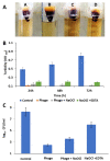
Irrigation efficiency of E. faecalis infected root canals. Panel (A) shows the two-chamber leakage model from root canals infected with E. faecalis isolate 4 and irrigated with either A. saline solution (control); B. phage treatment (vB_ZEFP 108 PFU/mL); C, phage plus 2.5% NaOCl; D. 2.5% NaOCl and 17% EDTA. Panel (B) shows culture turbidity measured every 24 h at OD600. Panel (C) shows viable counts after 72 h. Error bars indicate ± standard deviation of the mean.

References

    1. Dahl A., Bruun N.E. Enterococcus faecalis infective endocarditis: Focus on clinical aspects. Expert Rev. Cardiovasc. Ther. 2013;11:1247–1257. doi: 10.1586/14779072.2013.832482. - DOI - PubMed
    1. Molander A., Reit C., Dahlen G., Kvist T. Microbiological status of root-filled teeth with apical periodontitis. Int. Endod. J. 1998;31:1–7. doi: 10.1046/j.1365-2591.1998.t01-1-00111.x. - DOI - PubMed
    1. Wang Q.Q., Zhang C.F., Chu C.H., Zhu X.F. Prevalence of Enterococcus faecalis in saliva and filled root canals of teeth associated with apical periodontitis. Int. J. Oral Sci. 2012;4:19–23. doi: 10.1038/ijos.2012.17. - DOI - PMC - PubMed
    1. Siqueira J.F., Jr., Rocas I.N., Ricucci D., Hulsmann M. Causes and management of post-treatment apical periodontitis. Br. Dent. J. 2014;216:305–312. doi: 10.1038/sj.bdj.2014.200. - DOI - PubMed
    1. Tebruegge M., Pantazidou A., Clifford V., Gonis G., Ritz N., Connell T., Curtis N. The age-related risk of coexisting meningitis in children with urinary tract infection. PLoS ONE. 2011;6:e26576. doi: 10.1371/journal.pone.0026576. - DOI - PMC - PubMed

LinkOut - more resources