Review
. 2021 Mar 3;10(3):537.
doi: 10.3390/cells10030537.

Cell Death and Inflammation: The Role of Mitochondria in Health and Disease

Affiliations
Review

Cell Death and Inflammation: The Role of Mitochondria in Health and Disease

Anna Picca et al. Cells. .

Abstract

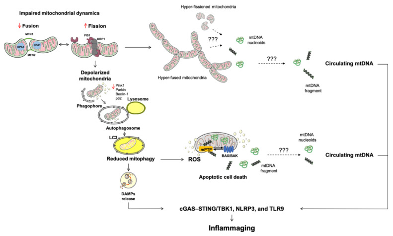
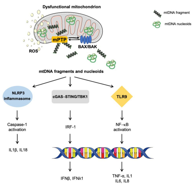
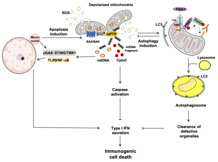
Mitochondria serve as a hub for a multitude of vital cellular processes. To ensure an efficient deployment of mitochondrial tasks, organelle homeostasis needs to be preserved. Mitochondrial quality control (MQC) mechanisms (i.e., mitochondrial dynamics, biogenesis, proteostasis, and autophagy) are in place to safeguard organelle integrity and functionality. Defective MQC has been reported in several conditions characterized by chronic low-grade inflammation. In this context, the displacement of mitochondrial components, including mitochondrial DNA (mtDNA), into the extracellular compartment is a possible factor eliciting an innate immune response. The presence of bacterial-like CpG islands in mtDNA makes this molecule recognized as a damaged-associated molecular pattern by the innate immune system. Following cell death-triggering stressors, mtDNA can be released from the cell and ignite inflammation via several pathways. Crosstalk between autophagy and apoptosis has emerged as a pivotal factor for the regulation of mtDNA release, cell's fate, and inflammation. The repression of mtDNA-mediated interferon production, a powerful driver of immunological cell death, is also regulated by autophagy-apoptosis crosstalk. Interferon production during mtDNA-mediated inflammation may be exploited for the elimination of dying cells and their conversion into elements driving anti-tumor immunity.

Keywords: apoptosis; damage-associated molecular patterns (DAMPs); immunogenic cell death; innate immunity; mitochondrial dynamics; mitochondrial dysfunction; mitochondrial quality control (MQC); mitophagy; oxidative stress; reactive oxygen species (ROS).


Conflict of interest statement

The authors declare no conflict of interest.

Figures

Figure 1
Figure 1
Schematic representation of altered mitochondrial quality control processes during aging and associated conditions. A multi-pathway derangement involving mitochondrial dynamics, mitophagy, and apoptosis characterizes mitochondrial dysfunction during aging and leads to cellular stress. Under these circumstances, the release of DAMPs ignite inflammation via innate immunity. Among other DAMPs, mtDNA is a strong pro-inflammatory molecule recognized by pattern recognition receptors. Of all mechanisms studied, the activation of BAK and BAX and the opening of the mitochondrial permeability transition pore are the best characterized route for the release of different molecules from mitochondria. Abbreviations: BAK, Bcl2 homologous antagonist/killer; BAX, BCL-associated X; cGAS–STING, GMP/AMP synthase—stimulator of interferon genes DNA-sensing system; DAMPs, damage-associated molecular patterns; DRP1, dynamin related protein 1; FIS1, mitochondrial fission 1 protein; MFN, mitofusin; mPTP, mitochondrial permeability transition pore; mtDNA, mitochondrial DNA; OPA1, optic atrophy protein 1; NLRP3, nucleotide-binding oligomerization domain-like receptor family pyrin domain containing 3; p62, sequestosome-1; PINK1, phosphatase and tensin homolog-induced kinase 1; ROS, reactive oxygen species; STING, stimulator of interferon genes protein; TBK1, TRAF family member-associated nuclear factor κB activator-binding kinase 1; TLR9, toll-like receptor 9.
Figure 2
Figure 2
Signaling pathways eliciting sterile inflammation via damaged-associated molecular patterns. In the setting of mitochondrial dysfunction and cellular stressful conditions, the activation of BAK and BAX and the opening of the mitochondrial permeability transition pore enable the mitochondrial release of a set of molecules collectively named damaged-associated molecular patterns. Among these, fragmented and/or oxidized mitochondrial DNA bound to the mitochondrial transcription factor A (green circles) can trigger inflammation via the activation of three main pro-inflammatory routes: (1) toll-like receptors, (2) nucleotide-binding oligomerization domain-like receptor family pyrin domain containing 3 inflammasome, and (3) cytosolic cyclic GMP/AMP synthase—stimulator of interferon genes DNA-sensing system. Abbreviations: BAK, Bcl2 homologous antagonist/killer; BAX, BCL-associated X; cGAS–STING, GMP/AMP synthase—stimulator of interferon genes DNA-sensing system; IFN, interferon; IL, interleukin; IRF-1, interferon regulatory factor 1; mPTP, mitochondrial permeability transition pore; mtDNA, mitochondrial DNA; NLRP3, nucleotide-binding oligomerization domain-like receptor family pyrin domain containing 3; NF-κB, nuclear factor κB; ROS, reactive oxygen species; TLR9, toll-like receptor 9; TBK1, TRAF family member-associated NF-κB activator-binding kinase 1; TNF-α, tumor necrosis factor alpha.
Figure 3
Figure 3
Schematic representation of apoptosis-driven caspase and autophagy activity eliciting immunogenic cell-death. Abbreviations: BAK, Bcl2 homologous antagonist/killer; BAX, BCL-associated X; cGAS–STING, GMP/AMP synthase—stimulator of interferon genes DNA-sensing system; CytoC, cytochrome C; IFN, interferon; LC3, microtubule-associated protein 1A/1B-light chain 3; mPTP, mitochondrial permeability transition pore; mtDNA, mitochondrial DNA; NF-κB, nuclear factor κB; PARL, presenilin-associated rhomboid-like protease; PINK1, phosphatase and tensin homolog-induced kinase 1; ROS, reactive oxygen species; TIM23, translocase of inner mitochondrial membrane 23; TLR9, toll-like receptor 9; TBK1, TRAF family member-associated NF-κB activator-binding kinase 1; TOM, translocase of the outer mitochondrial membrane.

References

    1. Ni H.M., Williams J.A., Ding W.X. Mitochondrial dynamics and mitochondrial quality control. Redox Biol. 2015;4:6–13. doi: 10.1016/j.redox.2014.11.006. - DOI - PMC - PubMed
    1. Picca A., Guerra F., Calvani R., Bucci C., Monaco M.R., Lo Bentivoglio A.R., Coelho-Júnior H.J., Landi F., Bernabei R., Marzetti E. Mitochondrial dysfunction and aging: Insights from the analysis of extracellular vesicles. Int. J. Mol. Sci. 2019;20:805. doi: 10.3390/ijms20040805. - DOI - PMC - PubMed
    1. López-Armada M.J., Riveiro-Naveira R.R., Vaamonde-García C., Valcárcel-Ares M.N. Mitochondrial dysfunction and the inflammatory response. Mitochondrion. 2013;13:106–118. doi: 10.1016/j.mito.2013.01.003. - DOI - PubMed
    1. Picca A., Lezza A.M.S., Leeuwenburgh C., Pesce V., Calvani R., Bossola M., Manes-Gravina E., Landi F., Bernabei R., Marzetti E. Circulating mitochondrial DNA at the crossroads of mitochondrial dysfunction and inflammation during aging and muscle wasting disorders. Rejuvenation Res. 2018;21:350–359. doi: 10.1089/rej.2017.1989. - DOI - PMC - PubMed
    1. Caielli S., Athale S., Domic B., Murat E., Chandra M., Banchereau R., Baisch J., Phelps K., Clayton S., Gong M., et al. Oxidized mitochondrial nucleoids released by neutrophils drive type I interferon production in human lupus. J. Exp. Med. 2016;213:697–713. doi: 10.1084/jem.20151876. - DOI - PMC - PubMed

Publication types

MeSH terms