Review
. 2021 Mar 17;22(6):3055.
doi: 10.3390/ijms22063055.

Factor XIII-A: An Indispensable "Factor" in Haemostasis and Wound Healing

Affiliations
Review

Factor XIII-A: An Indispensable "Factor" in Haemostasis and Wound Healing

Fahad S M Alshehri et al. Int J Mol Sci. .

Abstract

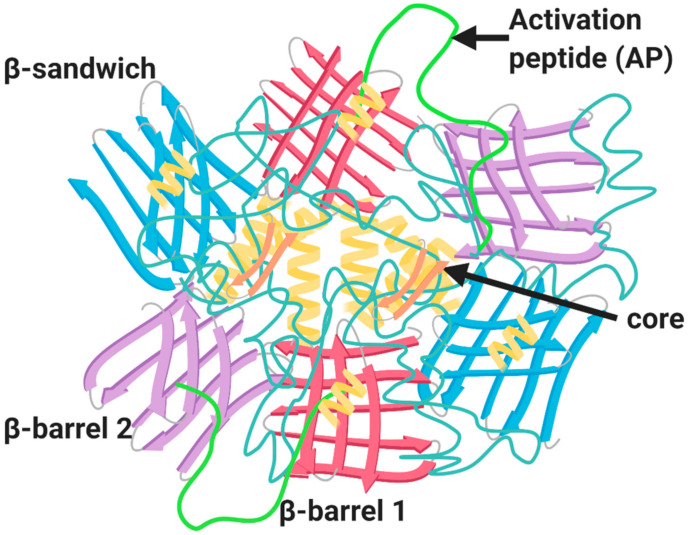
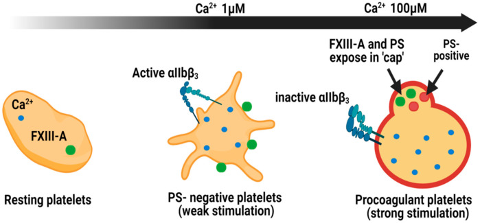
Factor XIII (FXIII) is a transglutaminase enzyme that catalyses the formation of ε-(γ-glutamyl)lysyl isopeptide bonds into protein substrates. The plasma form, FXIIIA2B2, has an established function in haemostasis, with fibrin being its principal substrate. A deficiency in FXIII manifests as a severe bleeding diathesis emphasising its crucial role in this pathway. The FXIII-A gene (F13A1) is expressed in cells of bone marrow and mesenchymal lineage. The cellular form, a homodimer of the A subunits denoted FXIII-A, was perceived to remain intracellular, due to the lack of a classical signal peptide for its release. It is now apparent that FXIII-A can be externalised from cells, by an as yet unknown mechanism. Thus, three pools of FXIII-A exist within the circulation: plasma where it circulates in complex with the inhibitory FXIII-B subunits, and the cellular form encased within platelets and monocytes/macrophages. The abundance of this transglutaminase in different forms and locations in the vasculature reflect the complex and crucial roles of this enzyme in physiological processes. Herein, we examine the significance of these pools of FXIII-A in different settings and the evidence to date to support their function in haemostasis and wound healing.

Keywords: Factor XIII-A; cellular FXIII-A; cross-linking; haemostasis; transglutaminase; wound healing.


Conflict of interest statement

The authors declare no conflict of interest.

Figures

Figure 1
Figure 1
Mechanisms of FXIII activation. (A) Thrombin- and Ca2+-driven cleavage of FXIIIA2B2. (B) Non-proteolytic activation of cellular FXIII-A by low Ca²⁺. The green and purple cylinders represent β-barrel and β-sandwich domains of FXIII-A subunit, respectively. The core domains in FXIII-A subunit are shown in orange and the activation peptides are shown in red. The inhibitory B subunits are shown in grey. Adapted from the work in [1].
Figure 2
Figure 2
Structure of FXIII-A zymogenic homodimer. The structure of FXIII-A homodimer is composed of four major domains: catalytic core domain (orange), β-sandwich domain (blue), β-barrel-1 domain (red) and the β-barrel-2 domain (purple); alpha helices strands (pale yellow); and the activation peptide (AP) (green).
Figure 3
Figure 3
Localisation of FXIII-A in platelet subpopulations. Within resting platelets, Ca2+ ions and cellular FXIII-A reside within the cytoplasm. Following weak stimulation, with agonists such as ADP, PS-negative platelets expose FXIII-A on the external membrane. The integrin αIIbβ3 is exposed in this subpopulation of platelets. Stimulation with dual agonists, such as thrombin and collagen, generate procoagulant platelets which expose FXIII-A within the phosphatidylserine (PS)-rich cap. In this subpopulation αIIbβ3 is inactive [70]. The cap is also rich in other coagulation factors, such as factor X, factor IX and prothrombin, and substrates for FXIII-A including fibrinogen, thrombospondin and factor Va [64].
Figure 4
Figure 4
The roles of Factor XIII in wound healing. During injury, FXIII-A enhances platelet adhesion to the injured endothelium through an integrin-dependent mechanism. Activated FXIII (FXIIIa) mediates the cross-linking of fibrin and promotes clot retraction. FXIII-A enhances invasion of monocytes/macrophages and phagocytosis of cell debris and pathogens. FXIII-A promotes crosslinking of extracellular matrix (ECM) and reduces vascular permeability. FXIIIa binds αvβ3 and promotes cross-linking to vascular endothelial growth factor receptor-2 (VEGFR-2) and downregulates thrombospondin-1 (TSP-1). Adapted from the work in [133].

References

    1. Bagoly Z., Koncz Z., Hársfalvi J., Muszbek L. Factor XIII, clot structure, thrombosis. Thromb. Res. 2012;129:382–387. doi: 10.1016/j.thromres.2011.11.040. - DOI - PubMed
    1. Lorand L. Factor XIII: Structure, Activation, and Interactions with Fibrinogen and Fibrin. Ann. N. Y. Acad. Sci. 2006;936:291–311. doi: 10.1111/j.1749-6632.2001.tb03516.x. - DOI - PubMed
    1. Karimi M., Bereczky Z., Cohan N., Muszbek L. Factor XIII Deficiency. Semin. Thromb. Hemost. 2009;35:426–438. doi: 10.1055/s-0029-1225765. - DOI - PubMed
    1. Yorifuji H., Anderson K., Lynch G.W., Van De Water L., McDonagh J. B protein of Factor XIII: Differentiation between free B and complexed B. Blood. 1988;72:1645–1650. doi: 10.1182/blood.V72.5.1645.1645. - DOI - PubMed
    1. Katona É., Pénzes D.K., Csapó A., Fazakas F., Udvardy M.L., Bagoly Z., Orosz Z.Z., Muszbek L. Interaction of Factor XIII subunits. Blood. 2014;123:1757–1763. doi: 10.1182/blood-2013-10-533596. - DOI - PubMed

LinkOut - more resources