Review
. 2021 Mar 17;22(6):3059.
doi: 10.3390/ijms22063059.

Therapeutic Role of Tocilizumab in SARS-CoV-2-Induced Cytokine Storm: Rationale and Current Evidence

Affiliations
Review

Therapeutic Role of Tocilizumab in SARS-CoV-2-Induced Cytokine Storm: Rationale and Current Evidence

Corrado Pelaia et al. Int J Mol Sci. .

Abstract

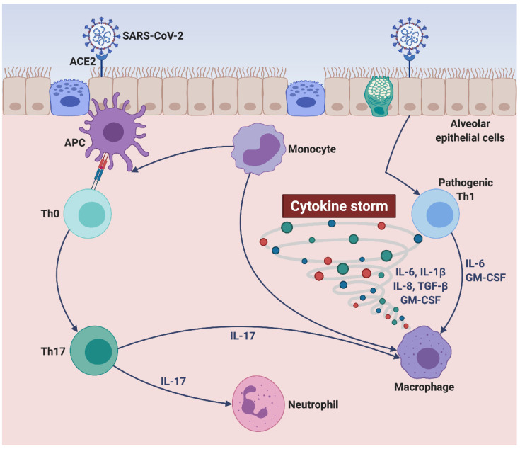
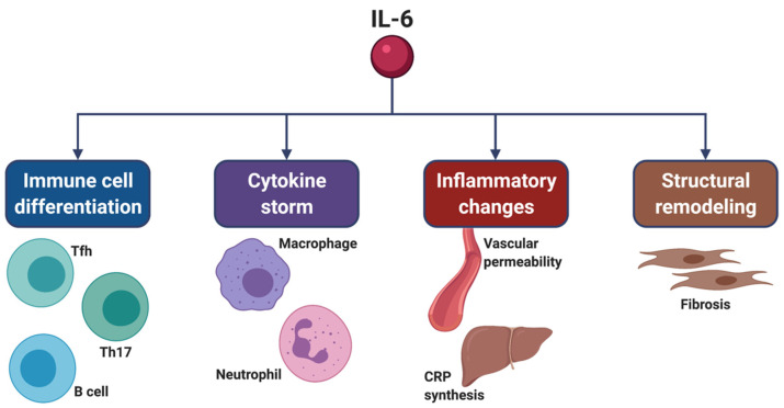
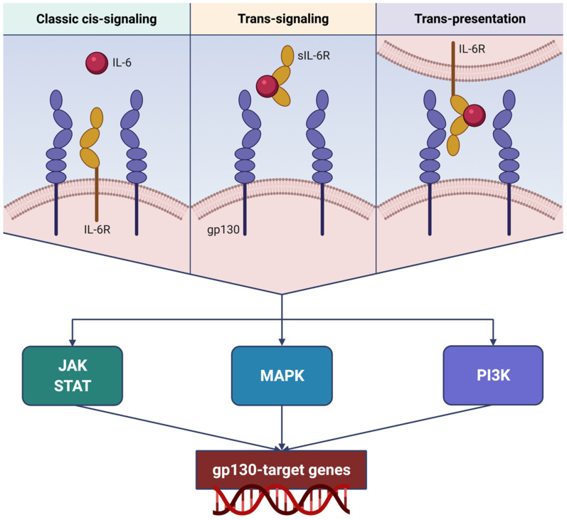
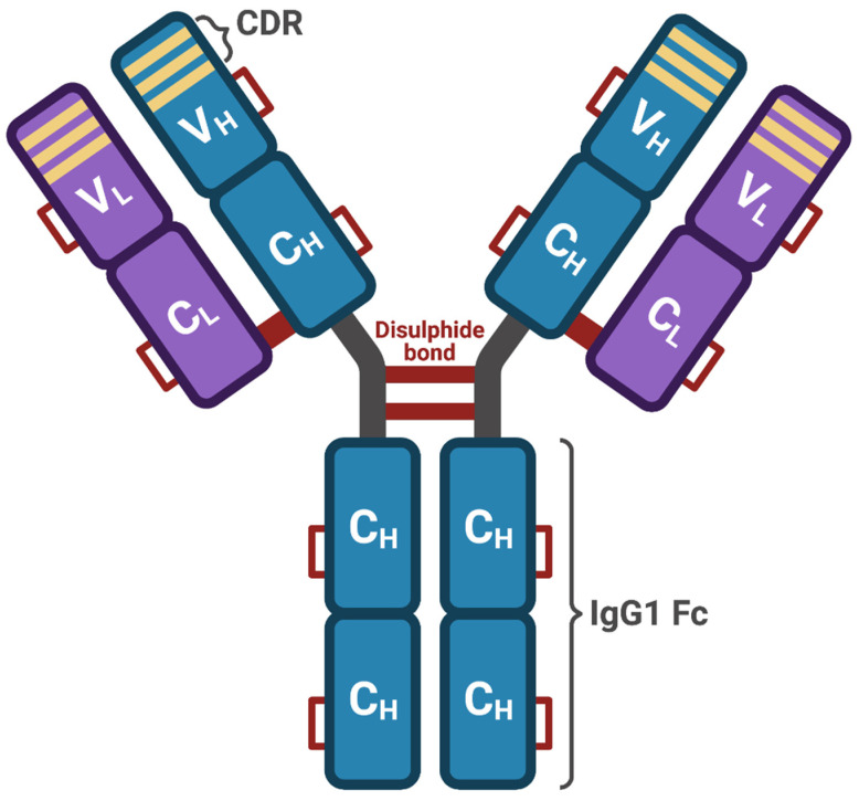
Among patients suffering from coronavirus disease 2019 (COVID-19) syndrome, one of the worst possible scenarios is represented by the critical lung damage caused by the severe acute respiratory syndrome coronavirus-2 (SARS-CoV-2)-induced cytokine storm, responsible for a potentially very dangerous hyperinflammatory condition. Within such a context, interleukin-6 (IL-6) plays a key pathogenic role, thus being a suitable therapeutic target. Indeed, the IL-6-receptor antagonist tocilizumab, already approved for treatment of refractory rheumatoid arthritis, is often used to treat patients with severe COVID-19 symptoms and lung involvement. Therefore, the aim of this review article is to focus on the rationale of tocilizumab utilization in the SARS-CoV-2-triggered cytokine storm, as well as to discuss current evidence and future perspectives, especially with regard to ongoing trials referring to the evaluation of tocilizumab's therapeutic effects in patients with life-threatening SARS-CoV-2 infection.

Keywords: ARDS; IL-6; SARS-CoV-2; cytokine storm; tocilizumab.


Conflict of interest statement

The authors declare that there is no conflict of interest.

Figures

Figure 1
Figure 1
SARS-CoV-2-induced cytokine storm. SARS-CoV-2 targets alveolar epithelial cells, binds to their ACE2 receptors, and penetrates inside lung tissue, thus reaching subepithelial antigen-presenting cells. The latter can drive the differentiation of unpolarized naïve T cells into mature Th17 lymphocytes producing IL-17, leading to neutrophil recruitment and macrophage activation. Moreover, upon their interactions with SARS-CoV-2, infected alveolar epithelial cells can trigger the activation of pathogenic Th1 cells and monocytes/macrophages, which release large quantities of proinflammatory cytokines (IL-6, GM-CSF, IL-1β, TGF-β) and chemokines (IL-8). Within such a cytokine/chemokine milieu (cytokine storm), monocytes can undergo further cellular differentiation towards either alveolar macrophage phenotype or antigen-presenting cell lineage. This original figure was created by the authors using BioRender.com. SARS-CoV-2: severe acute respiratory syndrome coronavirus-2; ACE2: angiotensin-converting enzyme 2; APC: antigen-presenting cell; Th0: unpolarized naïve T helper cell; Th1: T helper 1 cell; Th17: T helper 17 cell; IL-1β: interleukin-1β; IL-6: interleukin-6; IL-8: interleukin-8; IL-17: interleukin-17; GM-CSF: granulocyte-macrophage colony-stimulating factor; TGF-β: transforming growth factor-β.
Figure 2
Figure 2
Pleiotropic actions of IL-6, which targets many cell types and tissue districts. IL-6 promotes the differentiation of B cells, as well as of Th17 cells and Tfh cells. By contributing to the differentiation of Th17 cells, IL-6 also induces neutrophil recruitment and macrophage activation, which occur as relevant consequences of cytokine storm. Moreover, IL-6 increases vascular permeability and the hepatic synthesis of CRP, thus significantly contributing to the development and persistence of inflammation. IL-6 also induces tissue remodeling via stimulation of both fibroblast proliferation and production of extracellular matrix proteins. This original figure was created by the authors using BioRender.com. IL-6: interleukin-6; Th17: T helper 17 cell; Tfh: T follicular helper cells; CRP: C-reactive protein.
Figure 3
Figure 3
Different mechanisms underlying IL-6R activation and signaling. IL-6 can either interact with membrane-bound IL-6R (classic cis-signaling), or bind to sIL-6R (trans-signaling), or even be trans-presented from dendritic cells through their surface IL-6R to T lymphocytes (trans-presentation). Classic cis-signaling, trans-signaling, and trans-presentation converge on downstream transduction pathways, consisting of dimeric gp130-dependent activation of complex networks including JAK/STAT, MAPK, and PI3K signaling modules. Via these intracellular enzyme systems, IL-6R-triggered biological signals reach the nucleus and stimulate gp130 target genes involved in cell growth and proliferation. This original figure was created by the authors using BioRender.com. IL-6: interleukin-6; IL-6R: interleukin-6 receptor; sIL-6R: soluble interleukin-6 receptor; gp130: glycoprotein 130; JAK: Janus kinases; STAT: signal transducers and activators of transcription; MAPK: mitogen-activated protein kinases; PI3K: phosphoinositide 3-kinase.
Figure 4
Figure 4
Tocilizumab structure. Tocilizumab is a recombinant humanized IgG1κ antibody, consisting of two identical light chains, each one including a variable (VL) and a constant (CL) domain, coupled to two identical heavy chains, each one including a variable domain (VH) and three constant domains (CH). Overall, this antibody structure includes 12 intrachain and 4 interchain disulfide bonds. The variable portion of tocilizumab includes the complementarity-determining regions (CDR), which bind the IL-6 receptor. This original figure was created by the authors using BioRender.com.

Similar articles

Cited by

References

    1. Huang C., Wang Y., Li X., Ren L., Zhao J., Hu Y., Zhang L., Fan G., Xu J., Gu X., et al. Clinical features of patients infected with 2019 novel coronavirus in Wuhan, China. Lancet. 2020;395:497–506. doi: 10.1016/S0140-6736(20)30183-5. - DOI - PMC - PubMed
    1. Pelaia C., Tinello C., Vatrella A., De Sarro G., Pelaia G. Lung under attack by COVID-19-induced cytokine storm: Pathogenic mechanisms and therapeutic implications. Ther. Adv. Respir. Dis. 2020;14:1753466620933508. doi: 10.1177/1753466620933508. - DOI - PMC - PubMed
    1. Azkur A.K., Akdis M., Azkur D., Sokolowska M., van de Veen W., Brüggen M.C., O’Mahony L., Gao Y., Nadeau K., Akdis C.A. Immune response to SARS-CoV-2 and mechanisms of immunopathological changes in COVID-19. Allergy. 2020;75:1564–1581. doi: 10.1111/all.14364. - DOI - PMC - PubMed
    1. Yi Y., Lagniton P.N., Ye S., Li E., Xu R.H. COVID-19: What has been learned and to be learned about the novel coronavirus disease. Int. J. Biol. Sci. 2020;16:1753–1766. doi: 10.7150/ijbs.45134. - DOI - PMC - PubMed
    1. Peng Y., Mentzer A.J., Liu G., Yao X., Yin Z., Dong D., Dejnirattisai W., Rostron T., Supasa P., Liu C., et al. Broad and strong memory CD4+ and CD8+ T cells induced by SARS-CoV-2 in UK convalescent individuals following COVID-19. Nat. Immunol. 2020;21:1336–1345. doi: 10.1038/s41590-020-0782-6. - DOI - PMC - PubMed

MeSH terms

LinkOut - more resources