Review
. 2021 Mar 9;13(3):883.
doi: 10.3390/nu13030883.

Nutrient-Induced Cellular Mechanisms of Gut Hormone Secretion

Affiliations
Review

Nutrient-Induced Cellular Mechanisms of Gut Hormone Secretion

Van B Lu et al. Nutrients. .

Abstract

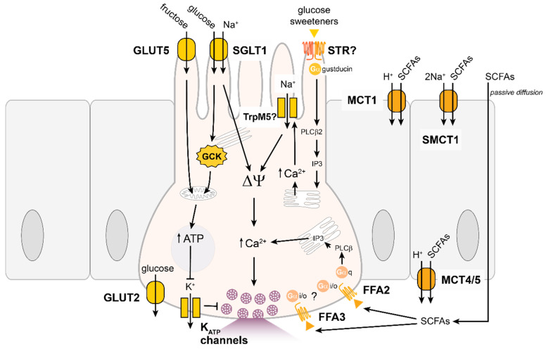
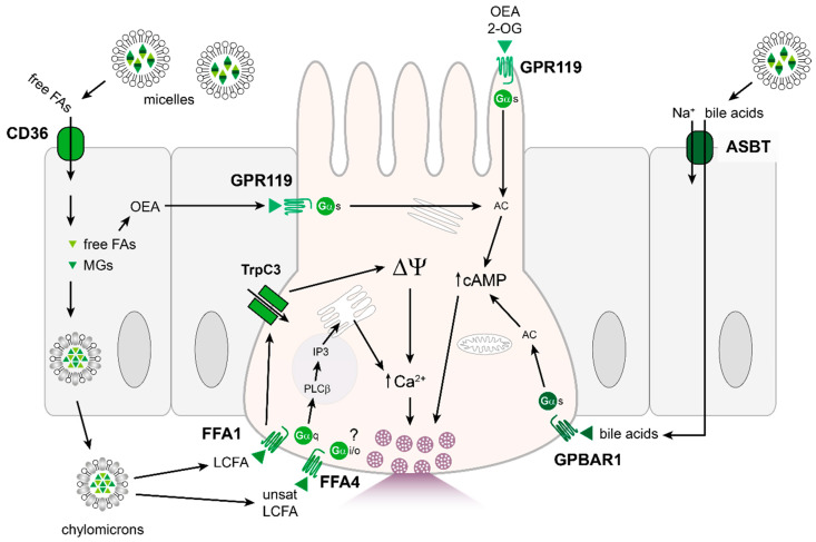
The gastrointestinal tract can assess the nutrient composition of ingested food. The nutrient-sensing mechanisms in specialised epithelial cells lining the gastrointestinal tract, the enteroendocrine cells, trigger the release of gut hormones that provide important local and central feedback signals to regulate nutrient utilisation and feeding behaviour. The evidence for nutrient-stimulated secretion of two of the most studied gut hormones, glucagon-like peptide 1 (GLP-1) and glucose-dependent insulinotropic polypeptide (GIP), along with the known cellular mechanisms in enteroendocrine cells recruited by nutrients, will be the focus of this review. The mechanisms involved range from electrogenic transporters, ion channel modulation and nutrient-activated G-protein coupled receptors that converge on the release machinery controlling hormone secretion. Elucidation of these mechanisms will provide much needed insight into postprandial physiology and identify tractable dietary approaches to potentially manage nutrition and satiety by altering the secreted gut hormone profile.

Keywords: GIP; GLP-1; chemosensory; enteroendocrine cells; hormones; nutrients.


Conflict of interest statement

The FR/FMG lab receives additional grant support from AstraZeneca and Eli Lilly for unrelated work. FMG is a consultant for Kallyope (New York, NY, USA).

Figures

Figure 1
Figure 1
Overview of macronutrient digestion. (A) Schematic representation of the distribution of glucose-dependent insulinotropic polypeptide (GIP) expressing K-cells and glucagon-like peptide 1 (GLP-1) expressing L-cells along the longitudinal intestinal axis. (B) Schematic representation of major sites of macronutrient digestion and absorption for carbohydrates, proteins and fats along the longitudinal intestinal axis. The breakdown of macronutrients denoted above primary location of nutrient absorption. Whereas absorbed monosaccharides and amino acids are exported from the intestinal epithelium as such, the majority of free fatty acids and monoglycerides are stepwise re-synthesised within the epithelium by MGAT2 and DGAT1 into triglycerides, which together with other lipophilic substances are secreted as chylomicrons. The production of lipid metabolites, such as OEA and 2-monoacylglycerides, which are synthesized following absorption of dietary fats, is represented in light green. Conjugated bile acids released following fat detection in the proximal small intestine is deconjugated in the distal intestine by colonic gut bacteria, as indicated by a dark green bar. Few macronutrients escape absorption in the small intestine, but bacterial fermentation of “indigestible fibres” provides SCFA as another nutritional source in the large intestine. Abbreviations: DGAT1, diacylglyceride-acyltransferase-1; GIP, glucose-dependent insulinotropic polypeptide; GLP-1, glucagon-like peptide 1; MGAT2, monoacylglyceride-acyltransferase-2; OEA, oleoylethanolamide; SCFAs, short-chain fatty acids.
Figure 2
Figure 2
Carbohydrate-sensing mechanisms in incretin hormone secreting EECs. Schematic of EEC with apical process (top) and basolateral surface (bottom) populated with secretory vesicles containing incretin hormones. Enterocytes shown in grey beside EEC. Glucose sensing by incretin-secreting EECs is critically dependent on sodium coupled uptake via SGLT1. Other sensors have been described, but are controversial (labelled with a question mark) or appear to be of limited physiological relevance. When SGLT1 is inhibited, GLP-1 secretion is elevated at later time points, possibly downstream of fermentation to short-chain fatty acids, which target G-protein coupled fatty acid receptors FFA2 and FFA3. These have been depicted on the basolateral membrane in analogy to the location of FFA1 (see text for details). Transport mechanisms of SCFAs across the intestinal epithelium illustrated in enterocytes on the right. Abbreviations: ATP, adenosine triphosphate; FFA2 or 3, Free fatty acid receptors; GCK, glucokinase; GLUT2, glucose transporter 2; GLUT5, glucose transporter 5; IP3, inositol triphosphate; KATP, ATP sensitive potassium channel; MCT1 or 4/5, monocarboxylate transporters; PLCβ, phospholipase C beta; SCFAs, short-chain fatty acids; SGLT1, sodium-glucose linked transporter 1; SMCT1, sodium-coupled monocarboxylate transporter 1; STR, sweet taste receptor; TrpM5, transient receptor potential cation channel subfamily M member 5; ΔΨ, membrane depolarization.
Figure 3
Figure 3
Protein-sensing mechanisms in incretin hormone secreting EECs. Schematic as in Fig2. Good evidence exists for a role of PEPT1, CaSR and GPR142 in incretin secretion, however, the exact locations of receptors and transporters are incompletely defined. Controversial contributors are marked with a question mark (see text for details). Abbreviations: AA, amino acid; B(0)AT-1, sodium-dependent neutral amino acid transporter; CaSR, calcium-sensing receptor; GPR142, G-protein coupled-receptor 142; GPRC6A, G protein-coupled receptor family C group 6 member A; IP3, inositol triphosphate; LPAR5, lysophosphatidic acid receptor 5; PEPT1, H+/peptide co-transporter; PLCβ, phospholipase C beta; TrpM5, transient receptor potential cation channel subfamily M member 5; UTR, umami taste receptor; ΔΨ, membrane depolarization.
Figure 4
Figure 4
Fat-sensing mechanisms in incretin hormone secreting EECs. Lipid sensing appears dominated by activation of G-protein coupled receptors. Contrary to what had been thought originally, these do not directly sample the luminal contents, but are shielded behind lipid absorption by enterocytes, with the possible exception of the mono-acyl-glyceride sensor GPR119. Absorption of micelles and formation of chylomicrons through enterocytes is illustrated on the left. Bile acids, which assist in the emulsification and breakdown of fatty acids, also stimulate incretin hormone secretion and the mechanism of bile acid-sensing in EECs is illustrated in dark green on right side of EEC. Transport route for conjugated bile acids in the small intestine via ASBT, illustrated in enterocyte on the right. See text for details. Abbreviations: 2-OG, 2-oleoylglycerol; AC, adenylyl cyclase; ASBT, sodium-dependent bile acid transporter; cAMP, cyclic adenosine monophosphate; CD36, cluster of differentiation also known as fatty acid translocase; FA; fatty acid; FFA1, free fatty acid receptor 1; FFA4, free fatty acid receptor 4; GPBAR1, G-protein coupled bile acid receptor 1; GPR119, G-protein coupled receptor 119; IP3, inositol triphosphate; LCFA, long-chain fatty acid; MG; monoglycerides; OEA, oleoylethanolamide; PLCβ, phospholipase C beta; TrpC3, transient receptor potential cation channel subfamily C member 3; ΔΨ, membrane depolarization.

References

    1. Bayliss W.M., Starling E.H. The mechanism of pancreatic secretion. J. Physiol. 1902;28:325–353. doi: 10.1113/jphysiol.1902.sp000920. - DOI - PMC - PubMed
    1. Bohórquez D.V., Chandra R., Samsa L.A., Vigna S.R., Liddle R.A. Characterization of basal pseudopod-like processes in ileal and colonic PYY cells. J. Mol. Histol. 2010;42:3–13. doi: 10.1007/s10735-010-9302-6. - DOI - PMC - PubMed
    1. Bohórquez D.V., Samsa L.A., Roholt A., Medicetty S., Chandra R., Liddle R.A. An Enteroendocrine cell—enteric glia connection revealed by 3D electron microscopy. PLoS ONE. 2014;9:e89881. doi: 10.1371/journal.pone.0089881. - DOI - PMC - PubMed
    1. McSwiney B.A., Spurrell W.R. Influence of osmotic pressure upon the emptying time of the stomach. J. Physiol. 1933;79:437–442. doi: 10.1113/jphysiol.1933.sp003057. - DOI - PMC - PubMed
    1. Knapper J., Heath A., Fletcher J., Morgan L., Marks V. GIP and GLP-1(7–36) amide secretion in response to intraduodenal infusions of nutrients in pigs. Comp. Biochem. Physiol. Part C Pharm. Toxicol. Endocrinol. 1995;111:445–450. doi: 10.1016/0742-8413(95)00046-1. - DOI - PubMed

MeSH terms

Substances

LinkOut - more resources