. 2021 Mar 9;13(3):882.
doi: 10.3390/nu13030882.

Soluble Fraction from Lysate of a High Concentration Multi-Strain Probiotic Formulation Inhibits TGF-β1-Induced Intestinal Fibrosis on CCD-18Co Cells

Affiliations

Soluble Fraction from Lysate of a High Concentration Multi-Strain Probiotic Formulation Inhibits TGF-β1-Induced Intestinal Fibrosis on CCD-18Co Cells

Francesca Lombardi et al. Nutrients. .

Abstract

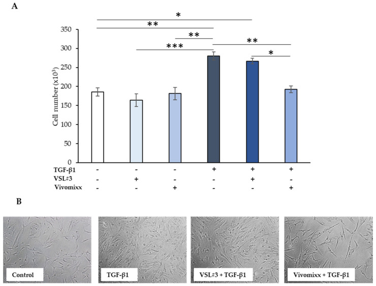
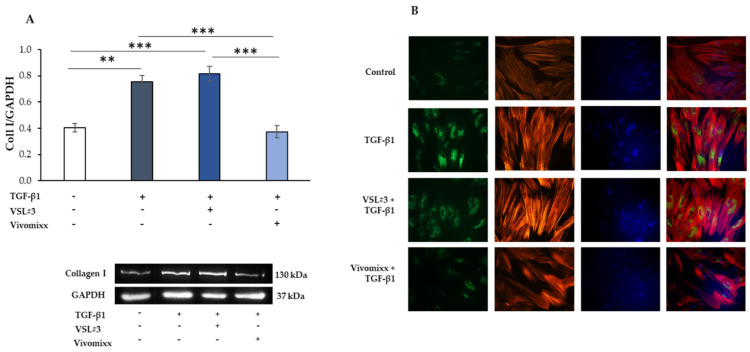
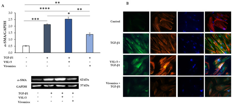
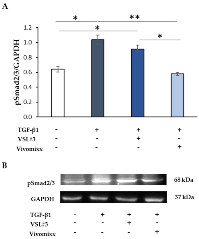
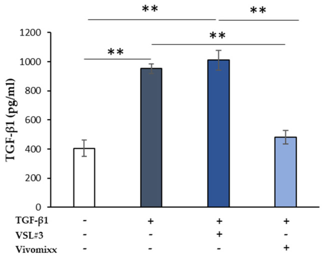
Fibrosis is a severe complication of chronic inflammatory disorders, such as inflammatory bowel disease (IBD). Current strategies are not fully effective in treating fibrosis; therefore, innovative anti-fibrotic approaches are urgently needed. TGF-β1 plays a central role in the fibrotic process by inducing myofibroblast differentiation and excessive extracellular matrix (ECM) protein deposition. Here, we explored the potential anti-fibrotic impact of two high concentration multi-strain probiotic formulations on TGF-β1-activated human intestinal colonic myofibroblast CCD-18Co. Human colonic fibroblast CCD-18Co cells were cultured in the presence of TGF-β1 to develop a fibrotic phenotype. Cell viability and growth were measured using the Trypan Blue dye exclusion test. The collagen-I, α-SMA, and pSmad2/3 expression levels were evaluated by Western blot analysis. Fibrosis markers were also analyzed by immunofluorescence and microscopy. The levels of TGF-β1 in the culture medium were assessed by ELISA. The effects of commercially available probiotic products VSL#3® and Vivomixx® were evaluated as the soluble fraction of bacterial lysates. The results suggested that the soluble fraction of Vivomixx® formulation, but not VSL#3®, was able to antagonize the pro-fibrotic effects of TGF-β1 on CCD-18Co cells, being able to prevent all of the cellular and molecular parameters that are related to the fibrotic phenotype. The mechanism underlying the observed effect appeared to be associated with inhibition of the TGF-β1/Smad signaling pathway. To our knowledge, this study provides the first experimental evidence that Vivomixx® could be considered to be a promising candidate against intestinal fibrosis, being able to antagonize TGF-β1 pro-fibrotic effects. The differences that were observed in our fibrosis model between the two probiotics used could be attributable to the different number of strains in different proportions.

Keywords: CCD-18Co cells; Smad 2/3; TGF-β1; VSL#3®; Vivomixx®; collagen-I; intestinal fibrosis; α-SMA.


Conflict of interest statement

The authors declare no conflict of interest.

Figures

Figure 1
Figure 1
Effect of the soluble fraction of bacterial lysates from VSL#3 and Vivomixx formulations on colonic intestinal fibroblast cells (CCD-18Co) cell number. (A) CCD-18Co cells were starved for 24 h and then incubated with TGF-β1 (10 ng/mL) for 48 h in the presence or absence of VSL#3- or Vivomixx-derived fractions (50 µg/mL), in serum-free medium. The values represent the cell number expressed as mean ± SEM of three independent experiments in duplicate. For comparative analysis of data groups, a one-way analysis of variance (ANOVA) with post hoc Bonferroni test was used. * p < 0.05, ** p < 0.01, *** p < 0.001. (B) Representative phase-contrast microscopic images showing CCD-18Co fibroblasts treated as described in (A).
Figure 2
Figure 2
Influence of the soluble fraction from VSL#3 and Vivomixx lysates on TGB-β1-induced collagen I expression in CCD-18Co cells. (A) Immunoblotting assay for collagen I was performed on CCD-18Co cells incubated for 48 h with TGF-β1 (10 ng/mL) in the presence or absence of VSL#3- or Vivomixx-derived fraction (50 µg/mL). Following densitometric analysis, obtained values were normalized vs. GAPDH. Data are from three independent experiments in duplicate, and values are expressed as mean ± SEM. For comparative analysis of data, a one-way analysis of variance (ANOVA) with post hoc Bonferroni test was used. ** p < 0.01, *** p < 0.001. A representative immunoblot of collagen I and GAPDH is also shown. (B) Representative immunofluorescence images of CCD-18Co cells treated as described in (A) and stained with anti-collagen I antibody (green) and with TRITC-phalloidin (red) to reveal F-actin. Nuclei were counterstained with DAPI (blue) (magnification 40×). The images are representative of three independent experiments in duplicate.
Figure 3
Figure 3
Influence of the soluble fraction from VSL#3 and Vivomixx lysates on TGB-β1-induced α-SMA expression in CCD-18Co cells. (A) Immunoblotting assay for α-SMA was performed on CCD-18Co cells incubated for 48 h with TGF-β1 (10 ng/mL) in the presence or absence of VSL#3- or Vivomixx-derived fraction (50 µg/mL). Following densitometric analysis, obtained values were normalized vs. GAPDH. Data are from three independent experiments in duplicate, and values are expressed as mean ± SEM. For comparative analysis of data, a one-way analysis of variance (ANOVA) with post hoc Bonferroni test was used. * p < 0.05, ** p < 0.01, *** p < 0.001, **** p < 0.0001. A representative immunoblot of α-SMA and GAPDH is also shown. (B) Representative immunofluorescence images of CCD-18Co cells treated, as described in A and stained with anti-α-SMA antibody (green) and with TRITC-phalloidin (red) to reveal F-actin. Nuclei were counterstained with DAPI (blue) (magnification 40×). The images are representative of three independent experiments in duplicate.
Figure 4
Figure 4
The effect of the soluble fraction from VSL#3 and Vivomixx lysates on TGF-β1-induced Smad activation. (A) Immunoblotting assay for pSmad2/3 protein expression was performed on CCD-18Co cells incubated for 48 h with TGF-β1 (10 ng/mL) in the presence or absence of VSL#3- or Vivomixx-derived fractions (50 µg/mL). Following densitometric analysis, the obtained values were normalized vs. GAPDH. The data are from two independent experiments in duplicate, and values are expressed as mean ± SEM. For comparative analysis of data, a one-way analysis of variance (ANOVA) with post hoc Bonferroni test was used. * p < 0.05, ** p < 0.01. (B) A Representative image of immunoblotting for pSmad2/3 and GAPDH is shown.
Figure 5
Figure 5
The effect of soluble fraction from VSL#3 or Vivomixx lysates on TGF-β1 secretion in activated CCD-18Co cells. TGF-β1 levels were detected by ELISA in supernatants from CCD-18Co cells that were treated for 48 h with TGF-β1 (10 ng/mL) in the presence or absence of VSL#3- or Vivomixx-derived fractions (50 µg/mL). The results, relative to one representative out of two experiments performed in triplicate, are expressed as mean ± SD. For comparative analysis of data groups, one-way ANOVA followed by post hoc test Bonferroni was used (** p < 0.01).

References

    1. Latella G., Di Gregorio J., Flati V., Rieder F., Lawrance I.C. Mechanisms of initiation and progression of intestinal fibrosis in IBD. Scand. J. Gastroenterol. 2015;50:53–65. doi: 10.3109/00365521.2014.968863. - DOI - PubMed
    1. Bettenworth D., Rieder F. Pathogenesis of Intestinal Fibrosis in Inflammatory Bowel Disease and Perspectives for Therapeutic Implication. Digest. Dis. 2017;35:25–31. doi: 10.1159/000449079. - DOI - PubMed
    1. Lawrance I.C., Rogler G., Bamias G., Breynaert C., Florholmen J., Pellino G., Reif S., Speca S., Latella G. Cellular and Molecular Mediators of Intestinal Fibrosis. J. Crohns Colitis. 2017;11:1491–1503. doi: 10.1016/j.crohns.2014.09.008. - DOI - PMC - PubMed
    1. Rieder F., Zimmermann E.M., Remzi F.H., Sandborn W.J. Crohn’s disease complicated by strictures: A systematic review. Gut. 2013;62:1072–1084. doi: 10.1136/gutjnl-2012-304353. - DOI - PMC - PubMed
    1. Latella G. Redox Imbalance in Intestinal Fibrosis: Beware of the TGFbeta-1, ROS, and Nrf2 Connection. Dig. Dis. Sci. 2018;63:312–320. doi: 10.1007/s10620-017-4887-1. - DOI - PubMed

MeSH terms

LinkOut - more resources