. 2021 Mar 9;11(3):185.
doi: 10.3390/membranes11030185.

The Early Fragmentation of a Bovine Dermis-Derived Collagen Barrier Membrane Contributes to Transmembraneous Vascularization-A Possible Paradigm Shift for Guided Bone Regeneration

Affiliations

The Early Fragmentation of a Bovine Dermis-Derived Collagen Barrier Membrane Contributes to Transmembraneous Vascularization-A Possible Paradigm Shift for Guided Bone Regeneration

Eleni Kapogianni et al. Membranes (Basel). .

Abstract

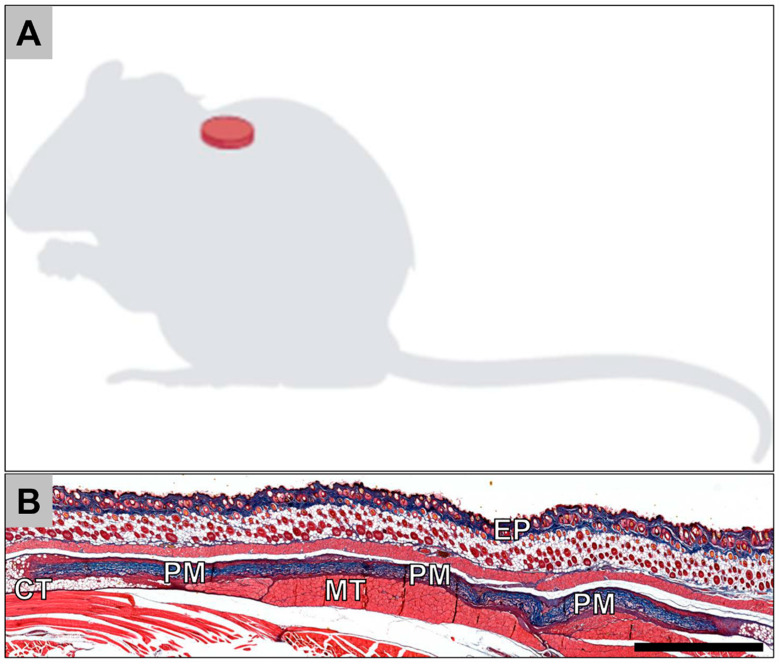
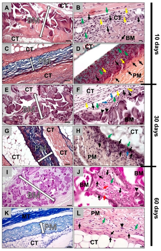
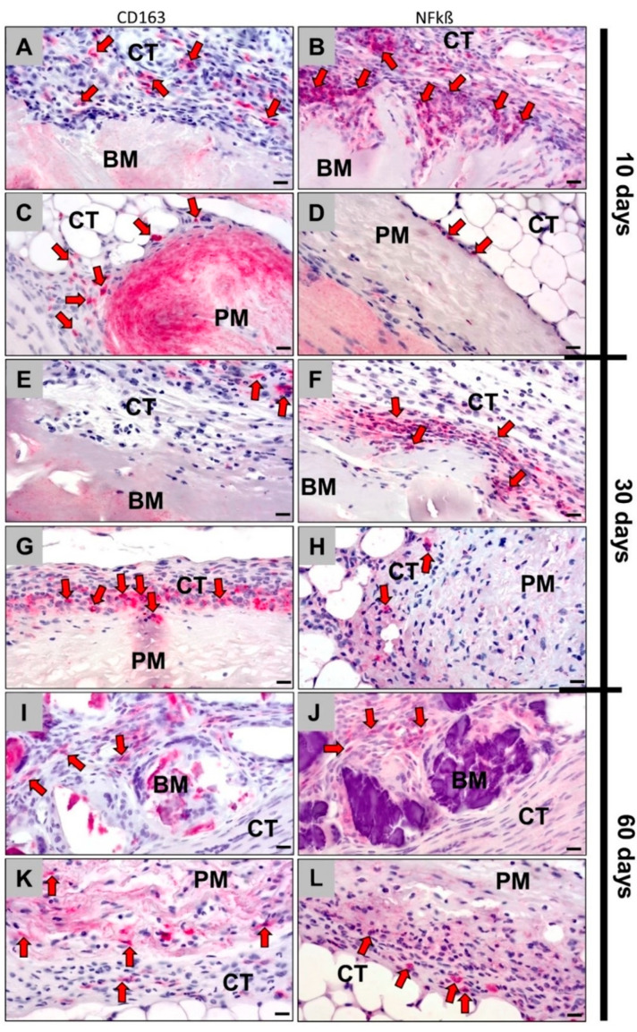
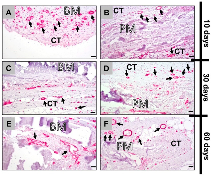
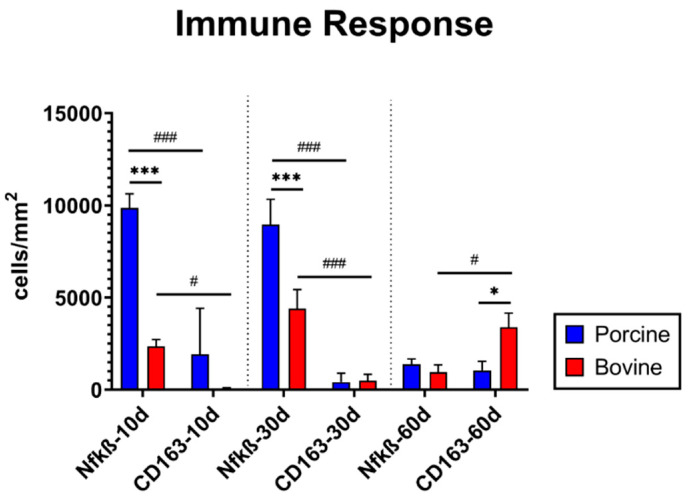
Collagen-based barrier membranes are an essential component in Guided Bone Regeneration (GBR) procedures. They act as cell-occlusive devices that should maintain a micromilieu where bone tissue can grow, which in turn provides a stable bed for prosthetic implantation. However, the standing time of collagen membranes has been a challenging area, as native membranes are often prematurely resorbed. Therefore, consolidation techniques, such as chemical cross-linking, have been used to enhance the structural integrity of the membranes, and by consequence, their standing time. However, these techniques have cytotoxic tendencies and can cause exaggerated inflammation and in turn, premature resorption, and material failures. However, tissues from different extraction sites and animals are variably cross-linked. For the present in vivo study, a new collagen membrane based on bovine dermis was extracted and compared to a commercially available porcine-sourced collagen membrane extracted from the pericardium. The membranes were implanted in Wistar rats for up to 60 days. The analyses included well-established histopathological and histomorphometrical methods, including histochemical and immunohistochemical staining procedures, to detect M1- and M2-macrophages as well as blood vessels. Initially, the results showed that both membranes remained intact up to day 30, while the bovine membrane was fragmented at day 60 with granulation tissue infiltrating the implantation beds. In contrast, the porcine membrane remained stable without signs of material-dependent inflammatory processes. Therefore, the bovine membrane showed a special integration pattern as the fragments were found to be overlapping, providing secondary porosity in combination with a transmembraneous vascularization. Altogether, the bovine membrane showed comparable results to the porcine control group in terms of biocompatibility and standing time. Moreover, blood vessels were found within the bovine membranes, which can potentially serve as an additional functionality of barrier membranes that conventional barrier membranes do not provide.

Keywords: Guided Bone Regeneration (GBR); barrier membrane; bovine collagen; bovine dermis; porcine collagen; porcine pericardium; tissue regeneration; tissue source; transmembraneous vascularization.


Conflict of interest statement

The authors declare no conflict of interest.

Figures

Figure 1
Figure 1
(A) Schematic image of the implantation site. (B) Overview of a subcutaneous implantation site of the porcine collagen membrane (PM) at day 30 postimplantation. CT = connective tissue; EP = epidermis (Azan-staining, “total scan”, 100× magnification, scalebar = 1 mm).
Figure 2
Figure 2
Histopathological images of bovine and porcine collagen membranes at days 10, 30 and 60. (A) Bovine collagen membrane at day 10 (hematoxylin and eosin (HE) staining, magnification = 200×, scale bar = 50 µm). (B) Bovine collagen membrane at day 10 (hematoxylin and eosin (HE) staining, magnification = 400×, scale bar = 20 µm). (C) Porcine collagen membrane at day 10 (Azan staining, magnification = 200×, scale bar = 50 µm). (D) Porcine collagen membrane at day 10 (Movat’s Pentachrome staining, magnification = 400×, scale bar = 20 µm). (E) Bovine collagen membrane at day 30 (hematoxylin and eosin (HE) staining, magnification = 200×, scale bar: 50 µm). (F) Bovine collagen membrane at day 30 (hematoxylin and eosin (HE) staining, magnification = 400×, scale bar = 20 µm). (G) Porcine collagen membrane at day 30 (Azan staining, magnification = 200×, scale bar = 50 µm). (H) Porcine collagen membrane at day 30 (hematoxylin and eosin (HE) staining, magnification = 400×, scale bar = 20 µm). (I) Bovine collagen membrane at day 60 (hematoxylin and eosin (HE) staining, magnification = 200×, scale bar = 50 µm). (J) Bovine collagen membrane at day 60 (hematoxylin and eosin (HE) staining, magnification = 400×, scale bar= 20 µm). (K) Porcine collagen membrane at day 60 (Giemsa staining, magnification = 200×, scale bar= 50 µm). (L) Porcine collagen membrane at day 60 (hematoxylin and eosin (HE) staining, magnification = 400×, scale bar = 20 µm). BM: bovine membrane; PM: porcine membrane; MT: muscle tissue; CT: connective tissue; white arrow: borders of the membrane; black arrows: macrophages; green arrows: fibroblasts; yellow arrows: eosinophils; blue arrows: neutrophils; black arrowheads: multinucleated giant cells; red arrows: blood vessels.
Figure 3
Figure 3
Immunohistochemically stained slides show detection of CD163-positive M2 macrophages (left column: A,C,E,G,I,K) and NF-kß-positive M1 macrophages (right column: B,D,F,H,J,L) into the implantation beds of both bovine and porcine collagen membranes at days 10, 30 and day 60 after implantation (all images: 400× magnification; scale bars = 20 μm) (left: CD163 immunohistochemical staining; right: NF-kß immunohistochemical staining). BM: bovine membrane; PM: porcine membrane; CT: connective tissue; red arrows: CD163- and NF-kß-positive macrophages.
Figure 4
Figure 4
Immunohistochemically stained slides show detection of CD31-positive endothelial cells into the implantation beds of both bovine (left column: A,C,E) and porcine collagen membranes (right column: B,D,F) at days 10, 30 and 60 after implantation (all images: 400× magnification; scalebars = 20 μm). CT: connective tissue; BM: bovine membrane; PM: porcine membrane; black arrows: CD31-positive vessels.
Figure 5
Figure 5
Results of the histomorphometrical analysis of the immune response within the implantation beds of both materials (*/# p ≤ 0.05, ***/### p ≤ 0.0001).

Similar articles

Cited by

References

    1. Tolstunov L., Hamrick J.F.E., Broumand V., Shilo D., Rachmiel A. Bone Augmentation Techniques for Horizontal and Vertical Alveolar Ridge Deficiency in Oral Implantology. Oral Maxillofac. Surg. Clin. N. Am. 2019;31:163–191. doi: 10.1016/j.coms.2019.01.005. - DOI - PubMed
    1. Omar O., Elgali I., Dahlin C., Thomsen P. Barrier membranes: More than the barrier effect? J. Clin. Periodontol. 2019;46:103–123. doi: 10.1111/jcpe.13068. - DOI - PMC - PubMed
    1. Elgali I., Omar O., Dahlin C., Thomsen P. Guided bone regeneration: Materials and biological mechanisms revisited. Eur. J. Oral Sci. 2017;125:315–337. doi: 10.1111/eos.12364. - DOI - PMC - PubMed
    1. Lee S.-W., Kim S.-G. Membranes for the Guided Bone Regeneration. Maxillofac. Plast. Reconstr. Surg. 2014;36:239–246. doi: 10.14402/jkamprs.2014.36.6.239. - DOI - PMC - PubMed
    1. Ghanaati S. In vivo Implantation of a Bovine-Derived Collagen Membrane Leads to Changes in the Physiological Cellular Pattern of Wound Healing by the Induction of Multinucleated Giant Cells: An Adverse Reaction? Front. Bioeng. Biotechnol. 2018;6:1–13. doi: 10.3389/fbioe.2018.00104. - DOI - PMC - PubMed

LinkOut - more resources