. 2021 Mar 9;11(3):217.
doi: 10.3390/life11030217.

Ferlins and TgDOC2 in Toxoplasma Microneme, Rhoptry and Dense Granule Secretion

Affiliations

Ferlins and TgDOC2 in Toxoplasma Microneme, Rhoptry and Dense Granule Secretion

Daniel N A Tagoe et al. Life (Basel). .

Abstract

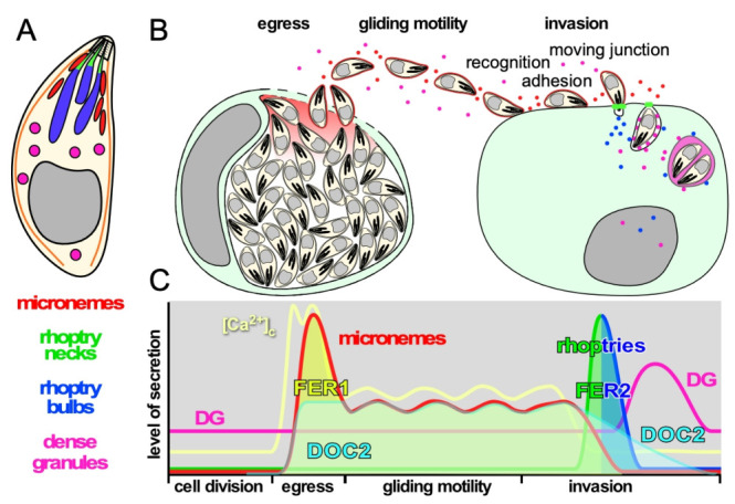
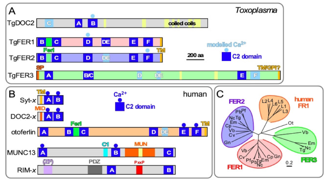
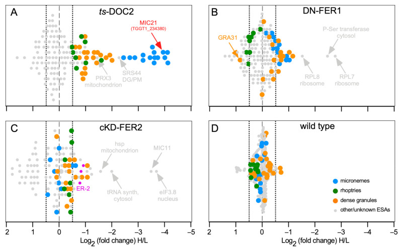
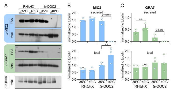
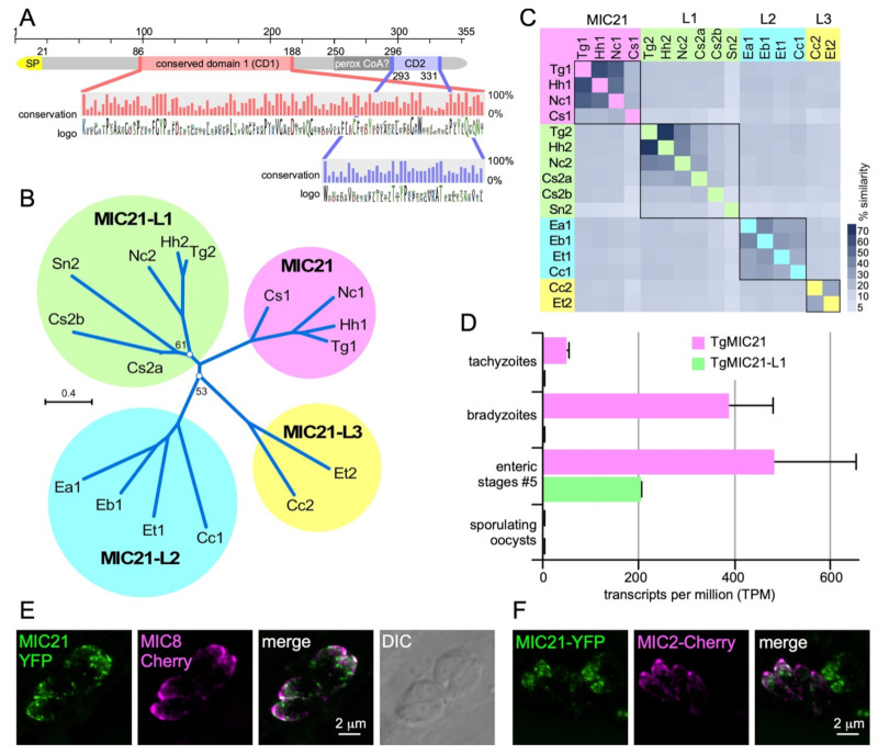
The host cell invasion process of apicomplexan parasites like Toxoplasma gondii is facilitated by sequential exocytosis of the microneme, rhoptry and dense granule organelles. Exocytosis is facilitated by a double C2 domain (DOC2) protein family. This class of C2 domains is derived from an ancestral calcium (Ca2+) binding archetype, although this feature is optional in extant C2 domains. DOC2 domains provide combinatorial power to the C2 domain, which is further enhanced in ferlins that harbor 5-7 C2 domains. Ca2+ conditionally engages the C2 domain with lipids, membranes, and/or proteins to facilitating vesicular trafficking and membrane fusion. The widely conserved T. gondii ferlins 1 (FER1) and 2 (FER2) are responsible for microneme and rhoptry exocytosis, respectively, whereas an unconventional TgDOC2 is essential for microneme exocytosis. The general role of ferlins in endolysosmal pathways is consistent with the repurposed apicomplexan endosomal pathways in lineage specific secretory organelles. Ferlins can facilitate membrane fusion without SNAREs, again pertinent to the Apicomplexa. How temporal raises in Ca2+ combined with spatiotemporally available membrane lipids and post-translational modifications mesh to facilitate sequential exocytosis events is discussed. In addition, new data on cross-talk between secretion events together with the identification of a new microneme protein, MIC21, is presented.

Keywords: Apicomplexa; DOC2; Toxoplasma; dense granule; exocytosis; microneme; rhoptry.


Conflict of interest statement

The authors declare no conflict of interest.

Figures

Figure 1
Figure 1
Schematic representation of the function and timing of secretion events in the T. gondii lytic cycle. (A). Schematic representation of T. gondii and its three secretory organelles, serving as legend for the other panels. (B). Schematic representation of secretion events throughout the lytic cycle. (C). Schematic representation of the levels of secretion of each organelle and the levels of intracellular calcium [Ca2+]c corresponding with the secretion events throughout the lytic cycle. Timeline on the X-axis corresponds with the events in panel B. Note that the invasion process completes in less than a minute and is shown in a stretched-out time window. The two spikes in [Ca2+]c at the time of egress correspond with the release of intracellular stores followed by the influx of extracellular Ca2+, while the [Ca2+]c oscillates during gliding in concert with bursts of motility [4]. The activity of DOC2 and FER1 and FER2 is also indicated.
Figure 2
Figure 2
Comparative topologies of Ca2+-mediated exocytosis machinery in Toxoplasma and humans and ferlin phylogeny. (A). Toxoplasma encodes four DOC2 domain proteins: TgDOC2 and three ferlin proteins. Ferlins are defined by 5 to 7 C2 domains. TgFER3 contains an N-terminal signal peptide (SP), which, in combination with the C-terminal TM domain, could signal GPI-anchor addition at the C-terminus. Yellow shades in TgFER3 represent coiled-coil domains. Light blue C2 domains are degenerate (defined as having a p-value below the cut-off in PFam database searches); light blue shaded Ca2+ binding to C2 is based on modeling. The C2 domains labeled A–F by established conventions [28,30,42]. TM: C-terminal transmembrane domain; FerI: conserved ferlin domain of unknown function. Modified from [43]. (B). The spectrum of types of C2 domain containing proteins in humans with roles in Ca2+-dependent secretion. Calcium sensors are present in the Syt-x, DOC-x, and ferlin (otoferlin shown as representative) families, though not all family members are Ca2+ sensors. MID = MUN Interaction Domain. MUN = Mammalian Uncoordinated domain. ZF = Zn Finger; PxxP = Pro-rich. (C). Phylogenetic analysis of apicomplexan, chromerid and human ferlins. The following abbreviations are used: human ferlins “L1-L5” FR1L1-5 (FR1L1 (dysferlin; O75923.1), FR1L2 (otoferlin; Q9HC10.3), FR1L3 (myoferlin; Q9NZM1.1), FR1L4 (A9Z1Z3.1), FR1L5 (A0AVI2.2), FR1L6 (Q2WGJ9.2)), Ot: green algae Ostreococcus tauri (Q01FJ7); Chromerids: “Vb” Vitrella brassicaformis (VbFER1 (Vbre_12074 + Vbra_12075), VbFER2 (Vbra_9198)) and “Cv” Chromera velia (CvFER1 (Cvel_17519.2) and CvFER2 (Cvel_9223)); Apicomplexa: “Tg” Toxoplasma gondii (TgFER1 (TGME49_309420), TgFER2 (TGME49_260470), TgFER3 (TGME49_295472 + TGME49_295468)) “Nc”, Neospora caninum (NcFER1 (NCLIV_053770), NcFER2 (NCLIV_026570), NcFER3 (NCLIV_002280)), “Em” Eimeria maxima (EmFER1 (EMWEY_00002120), EmFER2 (EMWEY_00009280), EmFER3 (EMWEY_00017650)), “Pf” Plasmodium falciparum (PfFER1 (PF3D7_0806300), PfFER2 (PF3D7_1455600)), “Pb” Plasmodium berghei (PbFER1 (PBANKA_122440), PbFER2 (PBANKA_131930)), “Cp” Cryptosporidium parvum (CpFER1 (cgd8_2910), CpFER2 (cgd2_2320)), “Gn” Gregarina niphandrodes (GnFER1 (GNI_063830), and GnFER2 (GNI_073830)). Alignment and unrooted Jukes-Cantor phylogenetic tree were generated in Geneious (v.6.1.6) [44]) from a MUSCLE alignment using neighbor-joining. Note that the FER1 and FER2 nodes for Tg and Nc are barely discernable at this scale. From [43].
Figure 3
Figure 3
SILAC data for T. gondii DOC2 and Ferlin mutants. (A). ts-DOC2 is a recreated temperature sensitive allele of DOC2 conditionally treated at 35 °C and 40 °C [43,47]; (B). DN-FER1 is a DD-Myc conditional overexpression allele induced with 1 μM Shield-1 [47]; (C). cKD-FER2 is tetracycline regulated promoter replacement induced with 1 μM ATc [59]; (D). the wild type control consisted of RHΔKu80 parasites treated at 35 °C and 40 °C. All inductions were performed for 48 hrs on intracellularly replicating parasites. For ESA collection, parasites were mechanically lysed from the host cell, and stimulated with 2% ethanol for 15 min. Parasites grown under permissive conditions were grown in light-isotope (L) labeled amino acids; parasites at restrictive conditions in heavy-isotope (H) labeled amino acids. Equal amounts of ESA collected under permissive and restrictive conditions were mixed and subjected to mass spectrometry. Averages of two biological repeats are shown, except for cKD-FER2 which was a single experiment. Marked in pink, the cKD-FER2 experiment is the only one in which some ER proteins (ER-2 pool defined in [117]) were detected (TGGT1_229480: putative calcium binding protein precursor; TGGT1_221210: cyclophilin; TGGT1_211680; protein disulfide isomerase). Dotted lines mark the −0.5 and +0.5 Log2 arbitrary cut-offs for decreased or increased significant changes, respectively, in excretion/secretion above background.
Figure 4
Figure 4
Dense granule secretion is TgDOC2 dependent. (A). Representative Western blots of ts-DOC2 secretion assays as performed for the ESA-SILAC experiment. “total” represents the pellet of the ESA assay. Antiserum against α-tubulin was used as loading control. Relative to the first lane for each condition, only half the protein amount was loaded in the second lane. (B). Quantification of secreted and total MIC2 normalized against α-tubulin shows that MIC2 expression is 3-fold upregulated in ts-DOC2 at the restrictive condition (40 °C), yet none is secreted. (C). Quantification of secreted and total GRA7 normalized against α-tubulin shows that GRA7 is not upregulated, but its stimulated secretion is dependent on DOC2. All blots used for quantification are provided in Supplementary Figure S2 and the quantification data in Supplementary Table S2. Statistics was performed with a two-tailed, two sample equal variance Student’s t-test; n.s.: non significant.
Figure 5
Figure 5
Characterization of putative microneme protein TGGT1_234380. TGGT1_234380 was the top hit in the ts-DOC2 SILAC experiment and annotated as a hypothetical protein on ToxoDB. Recently, the protein was detected in the microneme fraction by HyperLOPIT [117]. Querying select, representative apicomplexan and chromerid annotated genomes in EuPathDB identified only orthologs in the Coccidia. All except the T. gondii annotation of the predicted gene contained an N-terminal signal peptide (SP), which upon closer examination, was also present in the T. gondii gene if the gene was not spliced. This re-annotated version of the protein, which we named microneme protein 21 as this is the next available number in the microneme protein family (MIC21), was used to assemble a protein alignment that is the basis of data presented in panels A-C. (A). Schematic representation of MIC21. Two highly conserved domains (CD1 and CD2) were identified whose conservation across orthologs and paralogs is shown in the magnified panel as % conservation and amino acid logo plots. A PFam search identified domain defined in the peroxisomal acyl-CoA oxidase-II domains 3 and 4 (SCOP domain d1is2a3) with a non-significant e-value of 1, and furthermore this domain is not conserved across ortho- and para-logs. (B). Unrooted (neighbor-joining, Jukes-Cantor, CLC Workbench) phylogenetic tree of representative MIC21 related sequences identified in the Coccidia resolves into 4 branches of ortho- and paralogs (MIC21 cluster next to MIC21-like proteins MIC21-L1, MIC21-L2, and MIC21-L3 clusters). Bootstrap analysis (100x) supported all branches with over 70%, except for the two branch points as indicated. Hh: Hammondia hammondi; Nc: Neospora caninum; Cs: Cystoisospora suis; Sn: Sarcocystis neurona; Ea, Eb, Et: Eimeria acervulina, brunetti and tenella, respectively; Cc: Cyclospora cayetanensis. (C). Heatmap representing the percentage of sequence similarity across the DSP1 protein family. Percentage similarity based on ClustalOmega alignment. (D). RNAseq data available on ToxoDB reveals MIC21-L1 is not expressed in tachyzoite but only in the enteric stages. MIC21 is robustly expressed tachyzoites, bradyzoites and the enteric stages [120]. Neither MIC21 nor MIC21-L1 is expressed in sporulating oocysts [121]. Error bars represent standard error [120]. (E,F). Exogenous, transient expression of genomic DNA spanning the TgMIC21 promoter (1.4 kb) and coding sequence N-terminally fused to the YFP reporter co-transfected with a MIC8-mCherry (E) or MIC2-mCherry (F) reporter indicates MIC21 resided in the micronemes though does not perfectly co-localize with either MIC8 or MIC2.

Similar articles

Cited by

References

    1. Montoya J.G., Liesenfeld O. Toxoplasmosis. Lancet. 2004;363:1965–1976. doi: 10.1016/S0140-6736(04)16412-X. - DOI - PubMed
    1. Blader I.J., Coleman B.I., Chen C.T., Gubbels M.J. Lytic cycle of toxoplasma gondii: 15 years later. Annu. Rev. Microbiol. 2015;69:463–485. doi: 10.1146/annurev-micro-091014-104100. - DOI - PMC - PubMed
    1. Carruthers V.B., Sibley L.D. Sequential protein secretion from three distinct organelles of toxoplasma gondii accompanies invasion of human fibroblasts. Eur. J. Cell Biol. 1997;73:114–123. - PubMed
    1. Vella S.A., Moore C.A., Li Z.H., Hortua Triana M.A., Potapenko E., Moreno S.N.J. The role of potassium and host calcium signaling in Toxoplasma gondii egress. Cell Calcium. 2021;94:102337. doi: 10.1016/j.ceca.2020.102337. - DOI - PMC - PubMed
    1. Carruthers V.B., Tomley F.M. Microneme proteins in apicomplexans. Subcell. Biochem. 2008;47:33–45. - PMC - PubMed

LinkOut - more resources