Review
. 2021 Mar 9;11(3):481.
doi: 10.3390/diagnostics11030481.

Recent Advances in Understanding the Role of Autophagy in Paediatric Brain Tumours

Affiliations
Review

Recent Advances in Understanding the Role of Autophagy in Paediatric Brain Tumours

Francesca Gatto et al. Diagnostics (Basel). .

Abstract

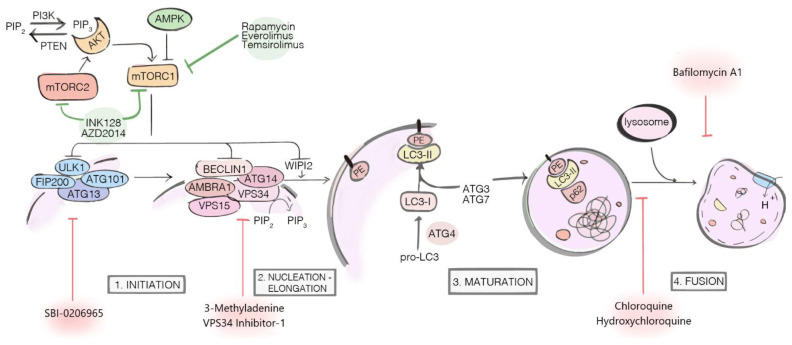
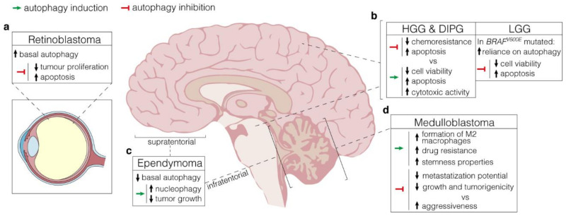
Autophagy is a degradative process occurring in eukaryotic cells to maintain homeostasis and cell survival. After stressful conditions including nutrient deprivation, hypoxia or drugs administration, autophagy is induced to counteract pathways that could lead to cell death. In cancer, autophagy plays a paradoxical role, acting both as tumour suppressor-by cleaning cells from damaged organelles and inhibiting inflammation or, alternatively, by promoting genomic stability and tumour adaptive response-or as a pro-survival mechanism to protect cells from stresses such as chemotherapy. Neural-derived paediatric solid tumours represent a variety of childhood cancers with unique anatomical location, cellular origins, and clinical presentation. These tumours are a leading cause of morbidity and mortality among children and new molecular diagnostics and therapies are necessary for longer survival and reduced morbidity. Here, we review advances in our understanding of how autophagy modulation exhibits antitumor properties in experimental models of paediatric brain tumours, i.e., medulloblastoma (MB), ependymoma (EPN), paediatric low-grade and high-grade gliomas (LGGs, HGGs), atypical teratoid/rhabdoid tumours (ATRTs), and retinoblastoma (RB). We also discuss clinical perspectives to consider how targeting autophagy may be relevant in these specific paediatric tumours.

Keywords: autophagy; brain tumours; oncology; targeted therapy.


Conflict of interest statement

The authors declare that the research was conducted in the absence of any commercial or financial relationships that could be construed as a potential conflict of interest.

Figures

Figure 1
Figure 1
Major components and regulators of the autophagy machinery. The autophagy inhibitors (red arrows) exert their actions at different stages of the process: SBI-0206965, 3-Methyladenine and VPS34 Inhibitor-1 are early-stage inhibitors, blocking autophagy before the autophagosome nucleation; Chloroquine (CQ), Hydroxychloroquine (HCQ) and Bafilomycin A1 are late-stage inhibitors, respectively blocking the autophagosome fusion with lysosome (CQ and HCQ) and inhibiting lysosomal acidification (Bafilomycin A1). The autophagy inducers (green arrows) Rapamycin, Everolimus, Temsirolimus, INK128, and AZD2014 target and inhibit mTOR1/2 complexes, indirectly activating autophagy.
Figure 2
Figure 2
Autophagy manipulation as a therapeutical strategy. (a). Retinoblastoma displays high protein expression of both LC3 and p62, which supports elevated basal levels of autophagy. Autophagy inhibition by lncRNA XIST targeting negatively affects tumour proliferation while increasing apoptosis; (b), LEFT, In HGG and DIPG, arising in the cerebral hemispheres and the pons respectively, autophagy manipulation results in controversial outcomes. Both activation (through the administration of a range of mTOR inhibitors) and inhibition (by 3-MA) have beneficial effects on cytotoxic activity and apoptosis, with detrimental consequences on chemoresistance. RIGHT, In BRAFV600E LGG, high autophagy levels have a pro-survival role. Hence, its inhibition through either pharmacologic or genetic approaches promotes apoptosis and reduces cell viability; (c), Supratentorial and infratentorial ependymomas are characterized by low basal levels of autophagy. Stimulation through rapamycin promotes nucleophagy and negatively affects tumour growth; (d), In medulloblastoma, autophagy induction via hypoxic conditions favours drug resistance and stemness properties through the formation of M2 macrophages in TME. Depending on the context, however, autophagy inhibition produces conflicting effects, either reducing metastasization potential and tumorigenicity or promoting aggressiveness.

Similar articles

Cited by

References

    1. Mizushima N., Komatsu M. Autophagy: Renovation of Cells and Tissues. Cell. 2011;147:728–741. doi: 10.1016/j.cell.2011.10.026. - DOI - PubMed
    1. Dikic I., Elazar Z. Mechanism and medical implications of mammalian autophagy. Nat. Rev. Mol. Cell Biol. 2018;19:349–364. doi: 10.1038/s41580-018-0003-4. - DOI - PubMed
    1. Yu L., Chen Y., Tooze S.A. Autophagy pathway: Cellular and molecular mechanisms. Autophagy. 2018;14:207–215. doi: 10.1080/15548627.2017.1378838. - DOI - PMC - PubMed
    1. Levine B., Kroemer G. Biological Functions of Autophagy Genes: A Disease Perspective. Cell. 2019;176:11–42. doi: 10.1016/j.cell.2018.09.048. - DOI - PMC - PubMed
    1. Ravanan P., Srikumar I.F., Talwar P. Autophagy: The spotlight for cellular stress responses. Life Sci. 2017;188:53–67. doi: 10.1016/j.lfs.2017.08.029. - DOI - PubMed

LinkOut - more resources