. 2021 Mar 9;22(5):2759.
doi: 10.3390/ijms22052759.

Increased Presence of Complement Factors and Mast Cells in Alveolar Bone and Tooth Resorption

Affiliations

Increased Presence of Complement Factors and Mast Cells in Alveolar Bone and Tooth Resorption

Kathrin Luntzer et al. Int J Mol Sci. .

Abstract

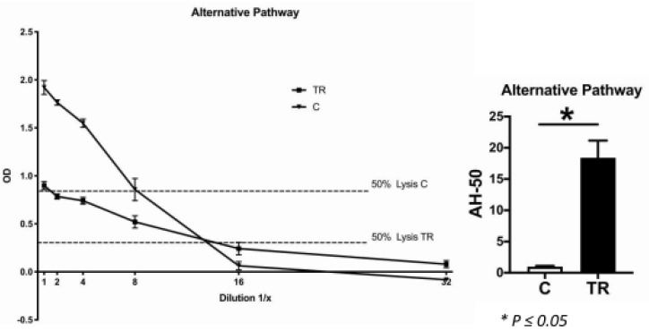
Periodontitis is the inflammatory destruction of the tooth-surrounding and -supporting tissue, resulting at worst in tooth loss. Another locally aggressive disease of the oral cavity is tooth resorption (TR). This is associated with the destruction of the dental mineralized tissue. However, the underlying pathomechanisms remain unknown. The complement system, as well as mast cells (MCs), are known to be involved in osteoclastogenesis and bone loss. The complement factors C3 and C5 were previously identified as key players in periodontal disease. Therefore, we hypothesize that complement factors and MCs might play a role in alveolar bone and tooth resorption. To investigate this, we used the cat as a model because of the naturally occurring high prevalence of both these disorders in this species. Teeth, gingiva samples and serum were collected from domestic cats, which had an appointment for dental treatment under anesthesia, as well as from healthy cats. Histological analyses, immunohistochemical staining and the CH-50 and AH-50 assays revealed increased numbers of osteoclasts and MCs, as well as complement activity in cats with TR. Calcifications score in the gingiva was highest in animals that suffer from TR. This indicates that MCs and the complement system are involved in the destruction of the mineralized tissue in this condition.

Keywords: complement system; mast cells; osteoclasts; periodontitis; tooth resorption.


Conflict of interest statement

The authors declare no conflict of interest.

Figures

Figure 1
Figure 1
(A) Von Kossa staining of the gingiva in control cats (C) (n = 4) and cats affected by gingivitis (G) (n = 6), periodontitis (PD) (n = 8) or tooth resorption (TR) (n = 11). Images were obtained at 100× magnification. In control and gingivitis sections, no crystals were observed (calcification score 0). In periodontitis samples, rare, tiny crystals, marked by black arrows, were present (calcification score 1). In this TR section many large crystals could be seen (calcification score 3). (B) There was a significant increase in the occurrence of crystals in the tooth resorption (TR) (n = 11) group compared with the control (C) (n = 4) and gingivitis (G) (n = 6) groups (both p ≤ 0.05). There was no significant difference between the periodontitis group (PD) (n = 8) and the other groups.
Figure 2
Figure 2
(A) Micro-Computed Tomography (μCT) image of a canine affected by tooth resorption (TR). The affected area is marked with a white rectangle. (B) Tartrate-resistant acid phosphatase staining of this tooth revealed many osteoclasts in this area, directly adjacent to the dentine of the tooth. The image was obtained at 400× magnification. (C) The evaluation showed significantly more osteoclasts in TR (n = 11) compared to periodontitis (PD) (n = 23) in both of the mineralized tissues, bone (p ≤ 0.01) and tooth (p ≤ 0.05). There was also a significant difference in the percentage of the eroded tooth surface between these groups (p ≤ 0.01).
Figure 3
Figure 3
(A) Image of the immunohistochemical histamine staining in a TR section. The image was taken at 200× magnification. MCs could be identified. (B) Immunohistochemical staining for histamine showed a significant increase of MCs in the TR group (n = 11) compared with the control (n = 7) and gingivitis groups (n = 10) (both p ≤ 0.01). There was no difference between the TR and periodontitis groups (n = 8).
Figure 4
Figure 4
(A) Immunohistochemical staining of the complement C5a receptor (C5aR). Images were taken at 400× magnification. The endothelial layer of the vessels in the gingiva of the controls (C) (n = 4) and cats affected by gingivitis (G) (n = 6), periodontitis (PD) (n = 7) or tooth resorption (TR) (n = 12) is marked by black arrows. In the controls and gingivitis animals, only a few endothelial layers were stained. The staining in general was less marked than in in the other two groups (PD, TR). (B) No significant differences were observed between the groups. However, there was a trend for a difference in periodontitis and TR compared to control and gingivitis regarding the endothelial layer. In addition, there was a trend for a difference in positively stained cells of the lamina propria between the control group and the other groups.
Figure 5
Figure 5
(A) Immunohistochemical staining of C5a in the gingiva of the controls (C) (n = 4) and of cats affected by gingivitis (G) (n = 6), periodontitis (PD) (n = 9) or tooth resorption (TR) (n = 12). Images were obtained at 400× magnification. No positive cells were observed in the control section. In the gingivitis section, a single positively stained cell was seen, marked by a black arrow. In the other two groups, notably more cells could be seen. (B) There was a trend for an increase in positively stained cells in the PD and TR groups compared with the other two groups. (C) Double immunofluorescence staining in the gingiva of a tooth affected by TR. Scale bar 50 μm. Mast cell granules (Avidin) were stained red, while C5a was stained green. Mast cells positive for C5a were observed, marked by white arrows.
Figure 6
Figure 6
(A) Immunohistochemical staining of complement factor C3 in the gingiva of the controls (C) (n = 4) and cats affected by gingivitis (G) (n = 6), periodontitis (PD) (n = 9) or tooth resorption (TR) (n = 12). Images were obtained at 400× magnification. In the control and gingivitis groups, no positively stained cells were observed. Two C3-positive cells were seen in periodontitis, marked by black arrows. The greatest number of positively stained cells could be observed in TR. (B) There was a statistically significant increase in C3-positive cells in the TR group compared with the other three groups (p ≤ 0.05). (C) Double immunofluorescence staining in the gingiva of a tooth affected by TR. Scale bar 50 μm. Mast cell granules (Avidin) were stained red, while C5 was stained green. In this section, one mast cell was positive for C3, marked by a white arrow.
Figure 7
Figure 7
Hemolytic assay for the detection of complement classical pathway function. The optical density of the serum samples of both the control (C) and tooth resorption (TR) groups were plotted against the dilution factor. The curves show the control (4 cats) and TR groups (12 cats) with different levels of complement function. In TR, 50% lysis occurred between serum dilutions 1:80 and 1:160, in the control group between the undiluted serum and dilution 1:10. There was a significant difference in the serum complement concentration between the two groups, being significantly higher in the TR group than in the control group (p ≤ 0.05).
Figure 8
Figure 8
Hemolytic assay for the detection of complement alternative pathway function. The optical density of the serum samples of both the control (C) and tooth resorption (TR) groups were plotted against the dilution factor. The curves show the control (4 cats) and TR groups (10 cats) with different levels of complement function. In both groups, 50% lysis occurred between serum dilutions 1:8 and 1:16. Nevertheless, there was a significant difference in the serum complement concentration between the two groups, being significantly higher in the TR group than in the control group (p ≤ 0.05).

Similar articles

Cited by

References

    1. Page R.C., Eke P.I. Case Definitions for Use in Population-Based Surveillance of Periodontitis. J. Periodontol. 2007;78:1387–1399. doi: 10.1902/jop.2007.060264. - DOI - PubMed
    1. Wolf H., Rateitschak-Pluss E., Rateitschak K.H. Color Atlas of Dental Medicine: Periodontology. Georg Thieme Verlag; Stuttgart, Germany: 2005.
    1. Silva N., Abusleme L., Bravo D., Dutzan N., Garcia-Sesnich J., Vernal R., Hernandez M., Gamonal J. Host response mechanisms in periodontal diseases. J. Appl. Oral Sci. Revista FOB. 2015;23:329–355. doi: 10.1590/1678-775720140259. - DOI - PMC - PubMed
    1. Shope B., Carle D. Tooth Resorptions in Dogs and Cats. [(accessed on 14 May 2017)]; Available online: http://blog.vetbloom.com/dentistry/tooth-resorption-in-dogs-and-cats/
    1. Weltman B., Vig K.W., Fields H.W., Shanker S., Kaizar E.E. Root resorption associated with orthodontic tooth movement: A systematic review. Am. J. Orthod. Dentofac. Orthop. 2010;137:462–476. doi: 10.1016/j.ajodo.2009.06.021. - DOI - PubMed

LinkOut - more resources