. 2021 Mar 9;13(3):439.
doi: 10.3390/v13030439.

Structure-Function Analyses of New SARS-CoV-2 Variants B.1.1.7, B.1.351 and B.1.1.28.1: Clinical, Diagnostic, Therapeutic and Public Health Implications

Affiliations

Structure-Function Analyses of New SARS-CoV-2 Variants B.1.1.7, B.1.351 and B.1.1.28.1: Clinical, Diagnostic, Therapeutic and Public Health Implications

Jasdeep Singh et al. Viruses. .

Abstract

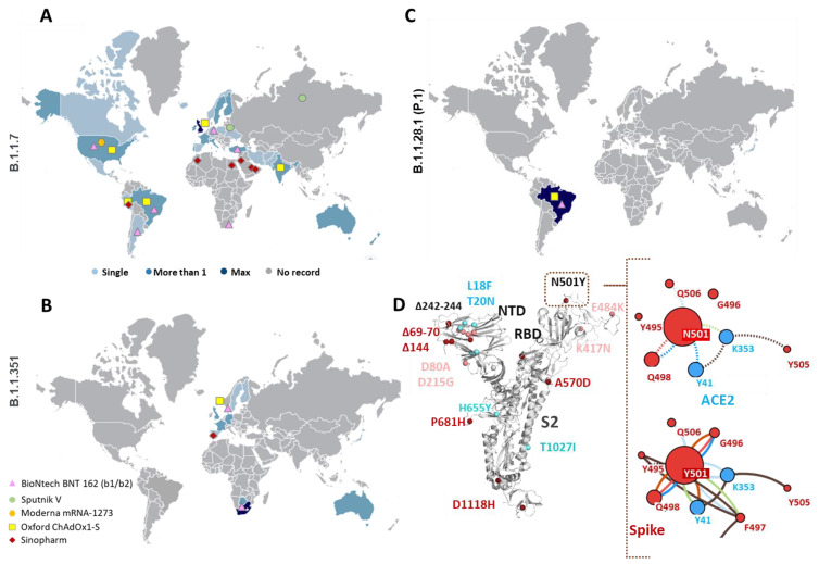
SARS-CoV-2 (Severe Acute Respiratory Syndrome-Coronavirus 2) has accumulated multiple mutations during its global circulation. Recently, three SARS-CoV-2 lineages, B.1.1.7 (501Y.V1), B.1.351 (501Y.V2) and B.1.1.28.1 (P.1), have emerged in the United Kingdom, South Africa and Brazil, respectively. Here, we have presented global viewpoint on implications of emerging SARS-CoV-2 variants based on structural-function impact of crucial mutations occurring in its spike (S), ORF8 and nucleocapsid (N) proteins. While the N501Y mutation was observed in all three lineages, the 501Y.V1 and P.1 accumulated a different set of mutations in the S protein. The missense mutational effects were predicted through a COVID-19 dedicated resource followed by atomistic molecular dynamics simulations. Current findings indicate that some mutations in the S protein might lead to higher affinity with host receptors and resistance against antibodies, but not all are due to different antibody binding (epitope) regions. Mutations may, however, result in diagnostic tests failures and possible interference with binding of newly identified anti-viral candidates against SARS-CoV-2, likely necessitating roll out of recurring "flu-like shots" annually for tackling COVID-19. The functional relevance of these mutations has been described in terms of modulation of host tropism, antibody resistance, diagnostic sensitivity and therapeutic candidates. Besides global economic losses, post-vaccine reinfections with emerging variants can have significant clinical, therapeutic and public health impacts.

Keywords: 501Y.V1; 501Y.V2; B.1.1.28.1; B.1.1.7; B.1.351; COVID-19 vaccines; Clade G; D614G variant; ORF8; P.1; furin cleavage site; immune escape; public health strategies; spike protein; vaccine delivery.


Conflict of interest statement

Authors declare no conflict of interest.

Figures

Figure 1
Figure 1
Emergence of new B.1.1.7, B.1.351 and P.1 variant lineages. (AC) Global distribution of sequences arising from various nations reporting 501Y.V1 (B.1.1.7), 501Y.V2 (B.1.351) and P.1 (B.1.1.28.1) variants, respectively. Pinned colored shapes on the map indicate major vaccine trials in various regions around the globe. Geographical pinning of vaccines in regions with rising frequency of variant population indicate the need to re-assess these candidates against new variants. Single (Light Blue), More than 1 (Blue) and Max (Dark Blue) indicate number of sequences of specific variants originating from different nations. (D) Structural mapping of mutations from 501Y.V1 (dark red dots) and 501Y.V2 (pink dots) variants on the spike (S) protein of Severe Acute Respiratory Syndrome-Coronavirus 2 (SARS-CoV-2). The amino acid deletions (Δ marked) in both variants are located in the N-terminal domain (NTD). The common N501Y mutation is located in receptor binding domain (RBD) region which makes contact with host angiotensin II converting enzyme (ACE2) receptors. Residue interactions of N501 (dashed lines) and Y501 (solid lines) with ACE2 (Blue dots) and other residues of the S protein (red dots) are shown in the right panel. Structural analysis of the mutations shows higher interaction network in Y501-ACE2 compared to wildtype N501-ACE2. Color codes—H-bonds (red), polar H-bonds (orange), VdW (light blue), aromatic (light green) and ring–ring interactions (brown).
Figure 2
Figure 2
Binding interactions and energy of Spike proteins with ACE-2. (A) The number of hydrogen bonds between the wildtype and mutant (N501Y and N440K) spike protein and ACE2 during the simulation. (B) Wildtype spike–ACE2 interactions in the average structure extracted from MD simulations, N501 (circled) making hydrophobic contact (hydrogen bonds are shown with green dots and non-polar interactions with magenta and brick semicircle). (C) N501Y–ACE2 interactions in the average structure. (D) Molecular mechanics energies combined with the generalized Born and surface area continuum solvation (MM/GBSA) binding free energy of spike proteins with ACE2.
Figure 3
Figure 3
Binding interactions and energy of spike proteins with C135 antibody. (A) The hydrogen bond peaks showing the interaction of mutants were better than wildtype spike protein. (B) The binding interaction of wildtype spike protein with heavy chain of C135 antibody, N501 (circled) making hydrophobic contact (hydrogen bonds are shown with green dots and non-polar interactions with magenta and brick semicircle). (C) The binding interaction of N501Y mutant spike protein with heavy chain of C135 antibody. (D) The binding interaction of N440K mutant spike protein with heavy chain of C135 antibody.
Figure 4
Figure 4
Binding interactions and energy of spike proteins with the CR3022 antibody. (A) The hydrogen interaction of wildtype was better than mutant spike proteins. (B) The binding interaction of wildtype spike protein with the heavy chain of CR3022 antibody, N501 (circled) making hydrophobic contact (hydrogen bonds are shown with green dots and non-polar interactions with magenta and brick semicircle). (C) The binding interaction of N501Y mutant spike protein with heavy chain of CR30222 antibody. (D) The binding interaction of N440K mutant spike protein with heavy chain of CR3022 antibody.
Figure 5
Figure 5
Binding affinity between ORF8 protomers. (A) The number of hydrogen bonds between the wildtype (WT) and mutant (MT) ORF8 protomers during the simulation. (B) Wildtype ORF8 interactions in the average structure extracted from MD simulations, R52 (circled) making hydrophobic contact (hydrogen bonds are shown with green dots and non-polar interactions with magenta and brick semicircle). (C) Mutant ORF8 interactions in the average structure. (D) MM/GBSA binding free energy in kcal/mol.

Similar articles

Cited by

References

    1. Sironi M., Hasnain S.E., Rosenthal B., Phan T., Luciani F., Shaw M.A., Sallum M.A., Mirhashemi M.E., Morand S., Gonzalez-Candelas F., et al. SARS-CoV-2 and COVID-19: A genetic, epidemiological, and evolutionary perspective. Infect. Genet. Evol. 2020;84:104384. doi: 10.1016/j.meegid.2020.104384. - DOI - PMC - PubMed
    1. Sheikh J.A., Singh J., Singh H., Jamal S., Khubaib M., Kohli S., Dobrindt U., Rahman S.A., Ehtesham N.Z., Hasnain S.E. Emerging genetic diversity among clinical isolates of SARS-CoV-2: Lessons for today. Infect. Genet. Evol. 2020;84:104330. doi: 10.1016/j.meegid.2020.104330. - DOI - PMC - PubMed
    1. Guan Q., Sadykov M., Mfarrej S., Hala S., Naeem R., Nugmanova R., Al-Omari A., Salih S., Mutair A.A., Carr M.J., et al. A genetic barcode of SARS-CoV-2 for monitoring global distribution of different clades during the COVID-19 pandemic. Int. J. Infect. Dis. 2020;100:216–223. doi: 10.1016/j.ijid.2020.08.052. - DOI - PMC - PubMed
    1. Singh H., Singh J., Khubaib M., Jamal S., Sheikh J.A., Kohli S., Hasnain S.E., Rahman S.A. Mapping the genomic landscape & diversity of COVID-19 based on >3950 clinical isolates of SARS-CoV-2: Likely origin & transmission dynamics of isolates sequenced in India. Indian J. Med. Res. 2020;151:474–478. doi: 10.4103/ijmr.IJMR_1253_20. - DOI - PMC - PubMed
    1. Korber B., Fischer W.M., Gnanakaran S., Yoon H., Theiler J., Abfalterer W., Hengartner N., Giorgi E.E., Bhattacharya T., Foley B., et al. Tracking changes in SARS-CoV-2 spike: Evidence that D614G increases infectivity of the COVID-19 virus. Cell. 2020;182:812–827.e19. doi: 10.1016/j.cell.2020.06.043. - DOI - PMC - PubMed

Publication types

MeSH terms

Substances