. 2021 Mar 9;13(3):441.
doi: 10.3390/v13030441.

Experimental Inoculation of Young Calves with SARS-CoV-2

Affiliations

Experimental Inoculation of Young Calves with SARS-CoV-2

Shollie Falkenberg et al. Viruses. .

Abstract

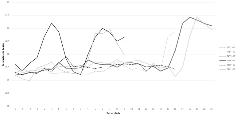
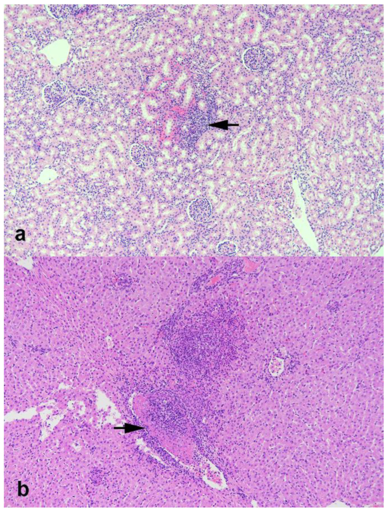
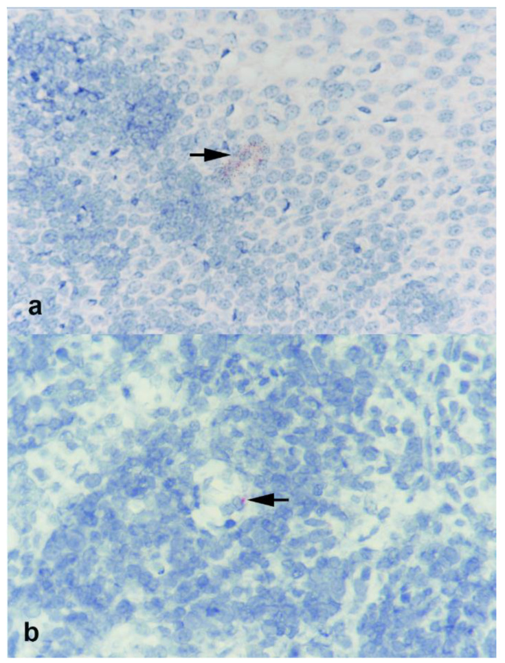
The host range of SARS-CoV-2 and the susceptibility of animal species to the virus are topics of great interest to the international scientific community. The angiotensin I converting enzyme 2 (ACE2) protein is the major receptor for the virus, and sequence and structural analysis of the protein has been performed to determine its cross-species conservation. Based on these analyses, cattle have been implicated as a potential susceptible species to SARS-CoV-2 and have been reported to have increased ACE2 receptor distribution in the liver and kidney, and lower levels in the lungs. The goal of the current study was to determine the susceptibility of cattle to SARS-CoV-2 utilizing inoculation routes that facilitated exposure to tissues with increased ACE2 receptor distribution. For this, colostrum-deprived calves approximately 6 weeks of age were inoculated via the intratracheal or intravenous routes. Nasal and rectal swab samples, as well as blood and urine samples, were collected over the course of the study to evaluate viral shedding, viremia, and seroconversion. Pyrexia was used as the primary criteria for euthanasia and tissue samples were collected during necropsy. Importantly, SARS-CoV-2 RNA was detected in only two nasal swab samples collected on days 3 and 10 post-inoculation (pi) in two calves; one calf in the intratracheal group and the other calf in the intravenous group, respectively. Additionally, the calf in the intratracheal group that was positive on the nasal swab on day 3 pi also had a positive tracheobronchial lymph node on day 9 pi. Viral nucleic acid load on these samples, based on PCR cycle threshold values, were low and infectious virus was not recovered from the samples. These results suggest that there was no productive replication of SARS-CoV-2 in calves following intratracheal and intravenous inoculation.

Keywords: SARS-CoV-2; bovine; inoculation.


Conflict of interest statement

The authors declare that they have no conflict of interests regarding the publication of this study.

Figures

Figure 1
Figure 1
Rumen temperature measurements over time for each respective calf.
Figure 2
Figure 2
Photomicrographs of kidney (a) and liver (b) from calves experimentally inoculated with SARS-CoV-2. (a) Focal area of hemorrhage and infiltrates of lymphocytes and lesser numbers of neutrophils (arrow). (b) Multifocal area of necrosis with infiltrates of mixed inflammatory cells (neutrophils, macrophages, lymphocytes). Note vasculitis with fibrinocellular thrombus (arrow). Hematoxylin and eosin (HE). Magnification = 10×.
Figure 3
Figure 3
Photomicrographs of (a) palatine tonsil and (b) tracheobronchial lymph nodes from calf 7441 experimentally inoculated with SARS-CoV-2 and examined 9 days later. Sparse labeling for viral RNA is noted at base of stratified squamous epithelial layer in tonsil and in lymphoid follicle of lymph node (arrows). RNAscope ISH SARS-CoV-2. Magnification = 40×.
Figure 4
Figure 4
Photomicrographs of (a) turbinate, (b) palatine tonsil, (c) lung, and (d) kidney from calves experimentally inoculated with SARS-CoV-2 and examined 9 days later. In both turbinate and tonsil, sparse labeling for ACE2 receptor is seen, associated with submucosal glands (arrows). Sparse labeling is also noted in lung (arrows) and within renal tubular epithelial cells in the kidney (arrows). BaseScope ISH ACE2. Magnification = 40X.

References

    1. Coronaviridae Study Group of the International Committee on Taxonomy of Viruses The species Severe acute respiratory syndrome-related coronavirus: Classifying 2019-nCoV and naming it SARS-CoV-2. Nat. Microbiol. 2020;5:536–544. doi: 10.1038/s41564-020-0695-z. - DOI - PMC - PubMed
    1. Zhu N., Zhang D., Wang W., Li X., Yang B., Song J., Zhao X., Huang B., Shi W., Lu R. A novel coronavirus from patients with pneumonia in China, 2019. N. Engl. J. Med. 2020 doi: 10.1056/NEJMoa2001017. - DOI - PMC - PubMed
    1. Zhou P., Yang X.-L., Wang X.-G., Hu B., Zhang L., Zhang W., Si H.-R., Zhu Y., Li B., Huang C.-L. A pneumonia outbreak associated with a new coronavirus of probable bat origin. Nature. 2020;579:270–273. doi: 10.1038/s41586-020-2012-7. - DOI - PMC - PubMed
    1. Wu F., Zhao S., Yu B., Chen Y.-M., Wang W., Song Z.-G., Hu Y., Tao Z.-W., Tian J.-H., Pei Y.-Y. A new coronavirus associated with human respiratory disease in China. Nature. 2020;579:265–269. doi: 10.1038/s41586-020-2008-3. - DOI - PMC - PubMed
    1. Andersen K.G., Rambaut A., Lipkin W.I., Holmes E.C., Garry R.F. The proximal origin of SARS-CoV-2. Nat. Med. 2020;26:450–452. doi: 10.1038/s41591-020-0820-9. - DOI - PMC - PubMed