. 2021 Mar 9;10(3):512.
doi: 10.3390/plants10030512.

Micromorphology, Ultrastructure and Histochemistry of Commelina benghalensis L. Leaves and Stems

Affiliations

Micromorphology, Ultrastructure and Histochemistry of Commelina benghalensis L. Leaves and Stems

Kareshma Doolabh et al. Plants (Basel). .

Abstract

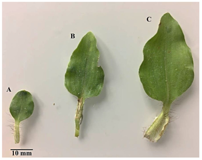
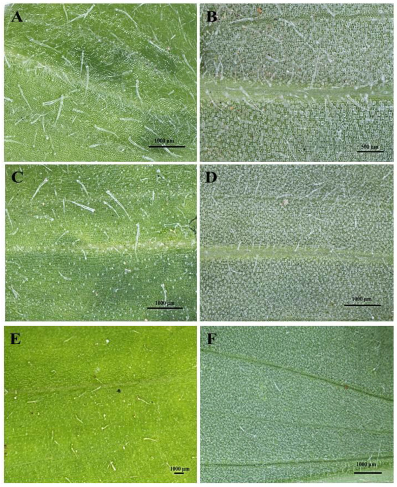
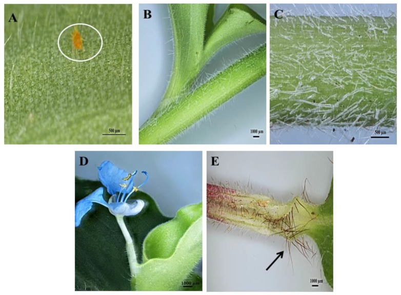
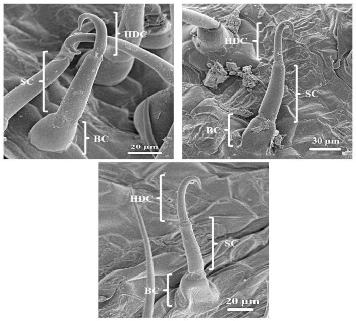
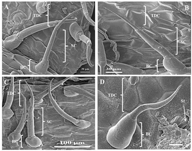
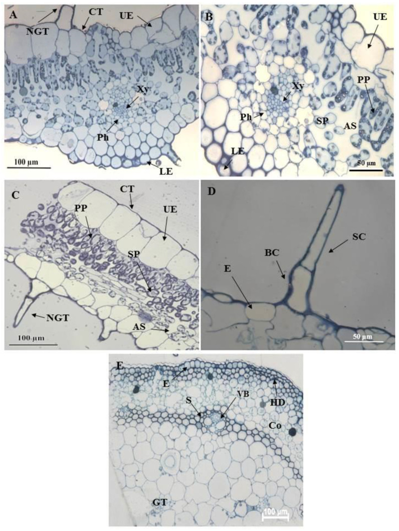
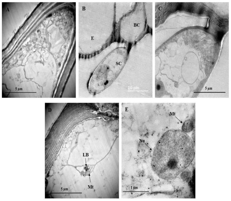
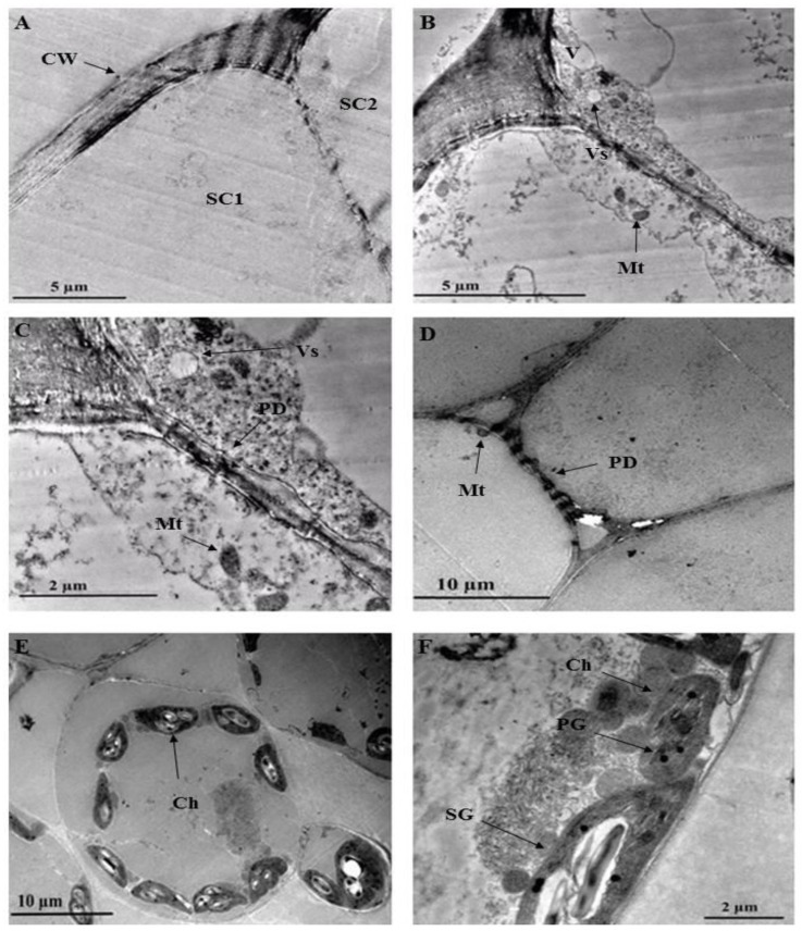
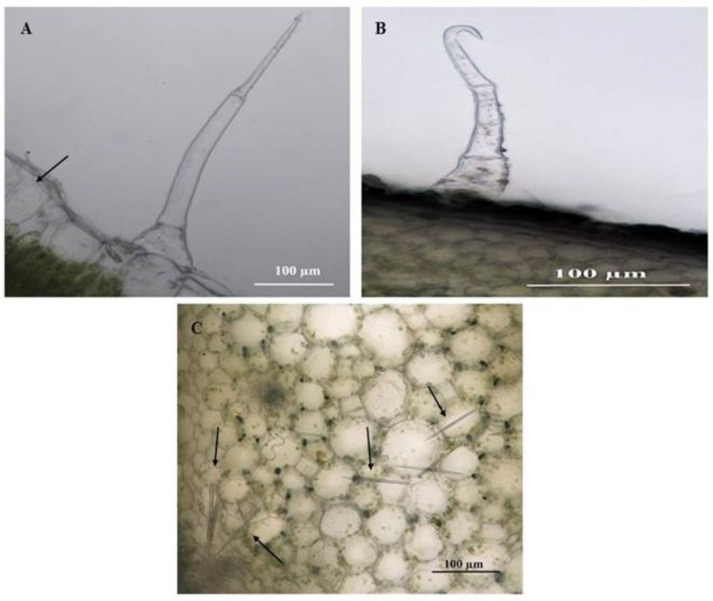
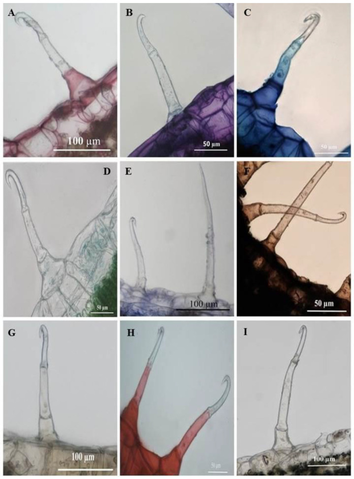
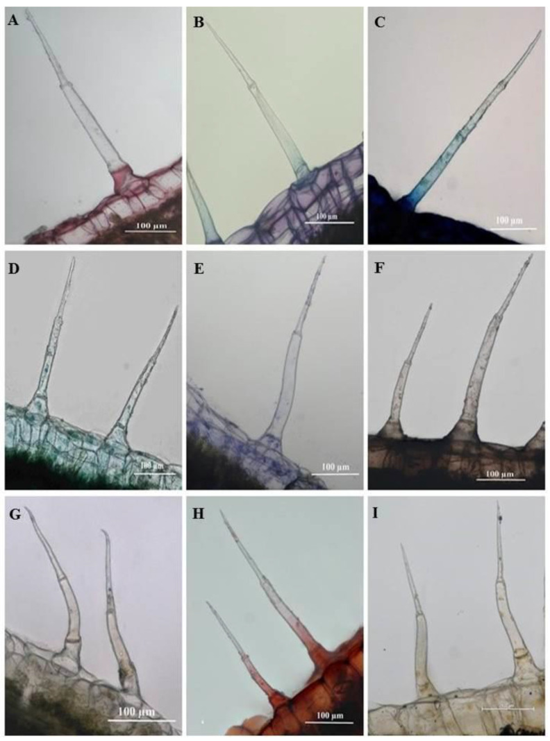
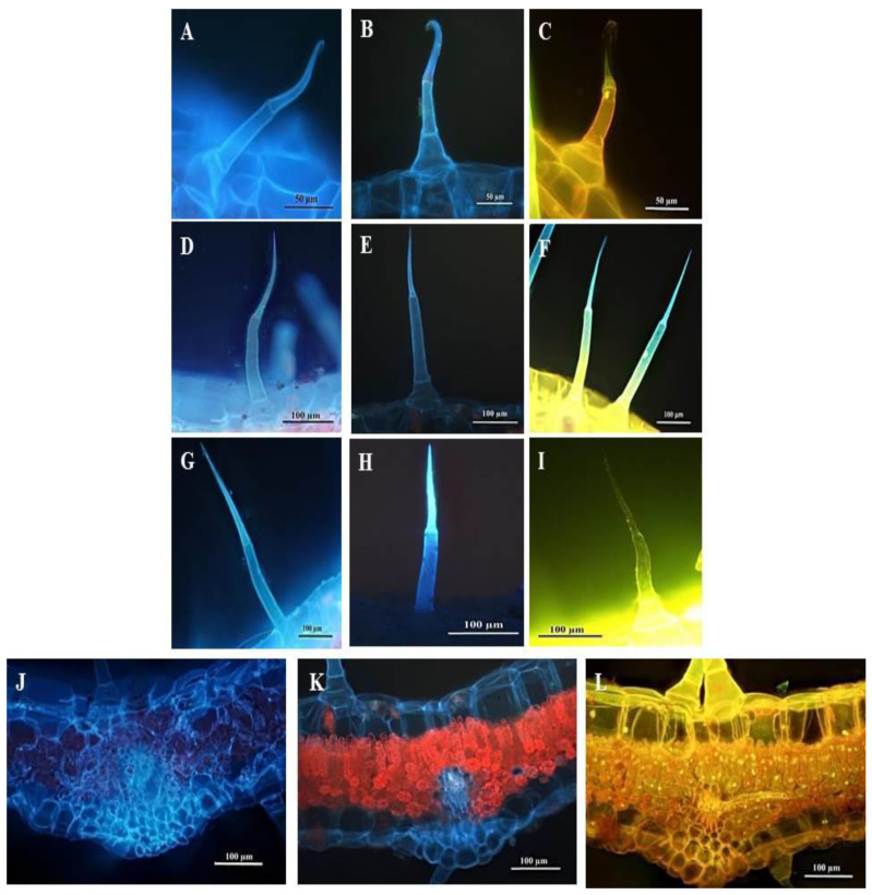
Commelina benghalensis L. is used as a traditional medicine in treating numerous ailments and diseases such as infertility in women, conjunctivitis, gonorrhea, and jaundice. This study used light and electron microscopy coupled with histochemistry to investigate the micromorphology, ultrastructure and histochemical properties of C. benghalensis leaves and stems. Stereo and scanning electron microscopy revealed dense non-glandular trichomes on the leaves and stems and trichome density was greater in emergent leaves than in the young and mature. Three morphologically different non-glandular trichomes were observed including simple multicellular, simple bicellular and simple multicellular hooked. The simple bicellular trichomes were less common than the multicellular and hooked. Transmission electron micrographs showed mitochondria, vesicles and vacuoles in the trichome. The leaf section contained chloroplasts with plastoglobuli and starch grains. Histochemical analysis revealed various pharmacologically important compounds such as phenols, alkaloids, proteins and polysaccharides. The micromorphological and ultrastructural investigations suggest that Commelina benghalensis L. is an economically important medicinal plant due to bioactive compounds present in the leaves and stems.

Keywords: alkaloids; hooked; microscopy; morphology; multicellular; non-glandular trichomes; phenols.


Conflict of interest statement

The authors declare no conflict of interest.

Figures

Figure 1
Figure 1
C. benghalensis leaves at different developmental stages. (A) Emergent; (B) Young; (C) Mature leaf.
Figure 2
Figure 2
Stereomicrographs of non-glandular trichomes on the adaxial and abaxial surfaces of C. benghalensis leaves. (A) Adaxial surface of emergent leaf; (B) Abaxial surface of emergent leaf; (C) Adaxial surface of young leaf; (D) Abaxial surface of young leaf; (E) Adaxial surface of mature leaf; (F) Abaxial surface of a mature leaf.
Figure 3
Figure 3
Stereomicrographs of non-glandular trichomes found on C. benghalensis plant parts. (A) Insect trapped among leaf trichomes; (B,C) Stem; (D) Flower and petiole; (E) Leaf sheath (red trichomes, arrow).
Figure 4
Figure 4
Scanning electron micrography of simple hooked non-glandular trichomes in all leaf stages and the stem of C. benghalensis. Abbreviations: BC: basal cell; SC: stalk cell; HDC: hooked distal cell.
Figure 5
Figure 5
Simple bicellular and multicellular non-glandular trichomes on leaves and stems of C. benghalensis. (A) Multicellular NGT on the midrib; (B,C) Multicellular NGT; (D) Bicellular NGT on the leaf and stem. Abbreviations: BC: basal cell; SC: stalk cell; TDC: tapered distal cell; St: stomata.
Figure 6
Figure 6
Light micrographs of resin embedded leaf and stem sections of C. benghalensis. (A,B) The midrib of an emergent leaf; (C) Segment of a leaf with a non-glandular trichome; (D) Non-glandular trichome on the epidermal layer of a leaf section; (E) Stem section Abbreviation: CT: cuticle; UE: upper epidermis; LE: lower epidermis; PP: palisade parenchyma; Xy: xylem; Ph: phloem; SP: spongy parenchyma; NGT: non-glandular trichome; BC: basal cell; SC: stalk cell; VB: vascular bundle; E: epidermis; HD: hypodermis; S: sclerenchyma; Co: collenchyma; GT: ground tissue; AS: air space.
Figure 7
Figure 7
Transmission electron micrograph of non-glandular trichomes found on C. benghalensis leaves. (A,B) Non-glandular trichomes on the leaf epidermis; (C) Cytoplasm containing electron dense material within the trichome; (D) Mitochondria and lamellar bodies located toward the periphery of the cell; (E) Nucleus and mitochondria within the cytoplasm. Abbreviations: BC: basal cell; SC: stalk cell; E: epidermis; LB: lamellar bodies; Mt: mitochondria; Nu: nucleus.
Figure 8
Figure 8
Transmission electron micrographs of C. benghalensis leaf section. (AC) Trichome; (DF) Leaf tissue containing chloroplasts and starch grains. Abbreviation: CW: cell wall; SC1: stalk cell 1; SC2: stalk cell 2; V: vacuole; Vs: vesicle; Mt: mitochondria; PD: plasmodesmata; Ch: chloroplast; PG: plastoglobuli; SG: starch grain.
Figure 9
Figure 9
Unstained leaf and stem sections of C. benghalensis. (A) Multicellular non-glandular trichome on a leaf that contains raphide crystals; (B) Hooked non-glandular trichome on the stem; (C) Raphide crystals inside stem tissue (Arrow).
Figure 10
Figure 10
Transverse sections of histochemically stained leaf sections of C. benghalensis. (A) Mucilage and pectin present in basal cells (Ruthenium red); (B) Toluidine blue for carboxylated polysaccharides (purple) and polyaromatic substances (blue); (C) Basal and stalk cell nucleoproteins (Methylene blue); (D) Cellulosic cell walls present in the basal cell (Fast green); (E) Coomassie blue stained proteins in the basal cell; (F) Lipids accumulated throughout the trichome (Sudan black); (G) Ferric chloride stained phenols in the basal and stalk cells (orange-brown); (H) Lignified cell walls in the basal and stalk cells stained red (Safranin); (I) Alkaloids were present in the basal and stalk cells stained with Wagner’s and Dittmar reagent (brown-orange).
Figure 11
Figure 11
Light micrographs of histochemically stained leaf sections of C. benghalensis. (A) Ruthenium Red stained basal cell red (mucilage and pectin); (B) Toluidine blue stained polyaromatic substances (blue) and carboxylated polysaccharides (purple); (C) Basal and stalk cell indicating nucleoproteins (Methylene blue); (D) Cellulosic cell walls in the basal cell indicated by Fast green; (E) Coomassie blue stained proteins in the basal cell; (F) Lipids accumulated throughout trichome (Sudan black); (G) Ferric chloride stained the basal and stalk cells orange-brown stain (phenols); (H) Lignified cell walls in the basal and stalk cells stained red (Safranin); (I) Alkaloids were present in the basal and stalk cells stained with Wagner’s and Dittmar reagent (brown-orange).
Figure 12
Figure 12
Fluorescent micrographs of leaf and stem sections of C. benghalensis. (A,D,G,J) Sections stained with Calcofluor white (cellulose); (B,E,H,K) Autofluorescence of sections (phenols); (C,F,I,L) Sections stained with Acridine orange (cell viability); (AC) Hooked trichomes on leaf sections; (DF) Multicellular trichomes on leaf sections; (GI) Multicellular trichomes on stem sections; (JL) Stained leaf sections.

Similar articles

References

    1. Hossain F., Saha S., Islam M.M., Nasrin S., Adhikari S. Analgesic and anti-inflammatory activity of Commelina benghalensis Linn. Turk. J. Pharm. Sci. 2014;11:25–32.
    1. Mbazima V.G., Mokgotho M.P., February F., Rees D.J.G., Mampuru L.J. Alteration of Bax-to-Bcl-2 ratio modulates the anticancer activity of methanolic extract of Commelina benghalensis (Commelinaceae) in Jurkat T cells. Afr. J. Biotechnol. 2008;7:3569–3576.
    1. Foden W., Potter L. Commelina benghalensis L. South African National Biodiversity Institute (SANBI) National Assessment: Red List of South African Plants Version 2017.1. [(accessed on 1 March 2021)];2005 Available online: http://redlist.sanbi.org/species.php?species=3576-13.
    1. Webster T.M., Burton M.G., Culpepper A.S., York A.C., Prostko E.P. Tropical spiderwort (Commelina benghalensis): A tropical invader threatens agroecosystems of the southern United States. Weed Technol. 2005;19:501–508. doi: 10.1614/WT-04-234R.1. - DOI
    1. Ghosh P., Dutta A., Biswas M., Biswas S., Hazra L., Nag S.K., Sil S., Chatterjee S. Phytomorphological, chemical and pharmacological discussions about Commelina benghalensis Linn. (Commelinaceae): A review. Pharma Innov. 2019;8:12–18.

LinkOut - more resources