Review
. 2021 Mar 18;8(3):39.
doi: 10.3390/bioengineering8030039.

Collagen-Based Electrospun Materials for Tissue Engineering: A Systematic Review

Affiliations
Review

Collagen-Based Electrospun Materials for Tissue Engineering: A Systematic Review

Britani N Blackstone et al. Bioengineering (Basel). .

Abstract

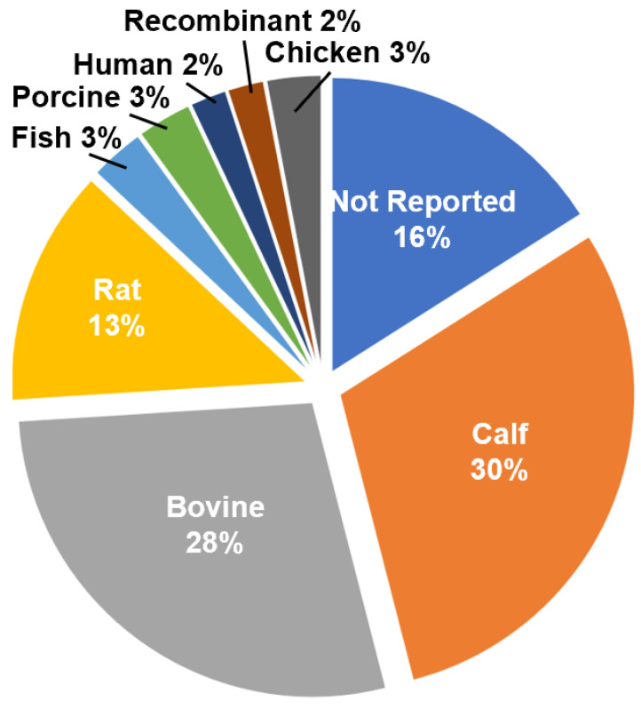
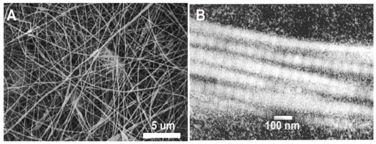
Collagen is a key component of the extracellular matrix (ECM) in organs and tissues throughout the body and is used for many tissue engineering applications. Electrospinning of collagen can produce scaffolds in a wide variety of shapes, fiber diameters and porosities to match that of the native ECM. This systematic review aims to pool data from available manuscripts on electrospun collagen and tissue engineering to provide insight into the connection between source material, solvent, crosslinking method and functional outcomes. D-banding was most often observed in electrospun collagen formed using collagen type I isolated from calfskin, often isolated within the laboratory, with short solution solubilization times. All physical and chemical methods of crosslinking utilized imparted resistance to degradation and increased strength. Cytotoxicity was observed at high concentrations of crosslinking agents and when abbreviated rinsing protocols were utilized. Collagen and collagen-based scaffolds were capable of forming engineered tissues in vitro and in vivo with high similarity to the native structures.

Keywords: collagen; electrospinning; tissue engineering.


Conflict of interest statement

The authors declare no conflict of interest.

Figures

Figure 1
Figure 1
Flow-through diagram for literature search and inclusion/exclusion criteria.
Figure 2
Figure 2
Distribution of collagen sources utilized in the study cohort for electrospinning.
Figure 3
Figure 3
(A) SEM image of a scaffold electrospun from calfskin collagen type I solubilized in 1,1,1,3,3,3-hexafluoro-2-propanol (HFP). (B) TEM image of an individual electrospun collagen fiber exhibiting D-banding. Modified from [22] with permission.
Figure 4
Figure 4
(A) Type of collagen-based electrospun scaffolds; (B) distribution of materials combined with collagen in electrospun scaffolds.
Figure 5
Figure 5
Confocal images of pure collagen (red) (A) and collagen (red)-polycaprolactone (PCL) (green) blend electrospun fibers (C) showing the segregation of the collagen and PCL components at a total PCL concentration of 30% total polymer mass. Scale bar = 7 µm. These large gelatin and PCL domains within the fiber reduced the ability of the collagen-PCL blend to support the formation of a well-stratified engineered skin (D) (blue = DAPI, red= collagen IV, green = involucrin) despite improvements to acellular scaffold strength and resistance to degradation. Pure collagen scaffolds support the development of bilayered engineered skin with a stratified epidermis (B). Modified from [75] with permission.

References

    1. Horbert V., Xin L., Foehr P., Brinkmann O., Bungartz M., Burgkart R.H., Graeve T., Kinne R.W. In vitro analysis of cartilage regeneration using a collagen type I hydrogel (CaReS) in the bovine cartilage punch model. Cartilage. 2018;10:346–363. doi: 10.1177/1947603518756985. - DOI - PMC - PubMed
    1. Hosseini Y., Verbridge S.S., Agah M. Bio-inspired microstructures in collagen type I hydrogel. J. Biomed. Mater. Res. Part A. 2014;103:2193–2197. doi: 10.1002/jbm.a.35352. - DOI - PubMed
    1. Boyce S.T. Fabrication, quality assurance, and assessment of cultured skin substitutes for treatment of skin wounds. Biochem. Eng. J. 2004;20:107–112. doi: 10.1016/j.bej.2003.09.017. - DOI
    1. Iejima D., Saito T., Uemura T. A collagen-phosphophoryn sponge as a scaffold for bone tissue engineering. J. Biomater. Sci. Polym. Ed. 2003;14:1097–1103. doi: 10.1163/156856203769231583. - DOI - PubMed
    1. Sumita Y., Honda M.J., Ohara T., Tsuchiya S., Sagara H., Kagami H., Ueda M. Performance of collagen sponge as a 3-D scaffold for tooth-tissue engineering. Biomaterials. 2006;27:3238–3248. doi: 10.1016/j.biomaterials.2006.01.055. - DOI - PubMed