. 2021 Mar 18;10(3):676.
doi: 10.3390/cells10030676.

Identification of Genetic Modifiers of TDP-43: Inflammatory Activation of Astrocytes for Neuroinflammation

Affiliations

Identification of Genetic Modifiers of TDP-43: Inflammatory Activation of Astrocytes for Neuroinflammation

Jae-Hong Kim et al. Cells. .

Abstract

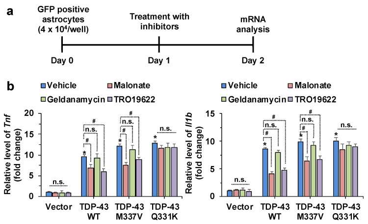
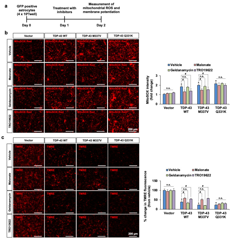
Transactive response DNA-binding protein 43 (TDP-43) is a ubiquitously expressed DNA/RNA-binding protein linked to amyotrophic lateral sclerosis (ALS) and frontotemporal dementia (FTD). TDP-43 has been implicated in numerous aspects of the mRNA life cycle, as well as in cell toxicity and neuroinflammation. In this study, we used the toxicity of the TDP-43 expression in Saccharomyces cerevisiae as an assay to identify TDP-43 genetic interactions. Specifically, we transformed human TDP-43 cDNAs of wild-type or disease-associated mutants (M337V and Q331K) en masse into 4653 homozygous diploid yeast deletion mutants and then used next-generation sequencing readouts of growth to identify yeast toxicity modifiers. Genetic interaction analysis provided a global view of TDP-43 pathways, some of which are known to be involved in cellular metabolic processes. Selected putative loci with the potential of genetic interactions with TDP-43 were assessed for associations with neurotoxicity and inflammatory activation of astrocytes. The pharmacological inhibition of succinate dehydrogenase flavoprotein subunit A (SDHA) and voltage-dependent anion-selective channel 3 (VDAC3) suppressed TDP-43-induced expression of proinflammatory cytokines in astrocytes, indicating the critical roles played by SDHA and VDAC3 in TDP-43 pathways during inflammatory activation of astrocytes and neuroinflammation. Thus, the findings of our TDP-43 genetic interaction screen provide a global landscape of TDP-43 pathways and may help improve our understanding of the roles of glia and neuroinflammation in ALS and FTD pathogenesis.

Keywords: TDP-43; amyotrophic lateral sclerosis; astrocyte; genetic interaction; glia; neuroinflammation.


Conflict of interest statement

The authors declare no conflict of interest.

Figures

Figure 1
Figure 1
Validation of toxicity suppressors by yeast spotting assays. Based on the analysis of Bar-seq data and Z-score distributions, the toxicity-suppressing yeast gene deletions were subjected to spotting assays. (ac) Overexpression of wild-type (WT) (a) or mutant human TDP-43 (b,c) causes toxicity in yeast. Deletion strains marked in blue show toxicity-suppressing effects. The pAG425GAL-ccdB yeast destination vector was used as the control. The BY4742 yeast strain was used as the WT control.
Figure 2
Figure 2
Interaction network of TDP-43. Human orthologs were identified for 13 yeast genes, the deletions of which suppressed the toxicity of TDP-43. A network view of the genetic interactions between TDP-43 and its interaction loci was generated using the GeneMANIA Cytoscape plugin. Other interacting or associated genes were also included in the network. TDP-43 genetic interactions are highlighted as thick blue lines (edges), and nodes are annotated according to biological processes.
Figure 3
Figure 3
The effects of pharmacological inhibition of SDHA, HSP90AB1, and VDAC3 on the inflammatory activation of astrocytes induced by TDP-43 transfection. (a) Astrocytes were transfected with a GFP, TDP-43 WT, TDP-43 M337V, or TDP-43 Q331K expression construct. After one day, FACS sorting of GFP-transfected cells was performed. (b) The relative levels of Tnf and Il1b mRNA in FACS-sorted primary astrocytes were assessed by real-time polymerase chain reaction (RT-PCR). Relative gene expression was normalized to the geometric mean of Gapdh and Actb. * p < 0.05 versus vehicle-treated control vector group; # p < 0.05 versus indicated groups; n.s., not significant. Data analysis by two-way analysis of variance (ANOVA) with four sister wells (biological replicates); mean ± standard deviation (SD).
Figure 4
Figure 4
The effects of pharmacological inhibition of SDHA, VDAC3, and HSP90AB1 on TDP-43 WT and mutant-induced mitochondrial dysfunction in primary astrocytes. (a) Experimental timeline. (b) Mitochondrial reactive oxygen species (ROS) was measured by MitoSOX staining. The fluorescence intensity of MitoSOX is shown as fold change in the adjacent graph. (c) Astrocytic mitochondrial membrane potential was assessed by TMRE probing. Percent reduction in TMRE fluorescence is shown in the adjacent graph. Scale bars indicate 200 µm. * p < 0.05 versus vehicle-treated control vector group; # p < 0.05 versus indicated groups; n.s., not significant. Data analysis by two-way ANOVA with four sister wells (biological replicates); mean ± SD.

References

    1. Buratti E., Baralle F.E. TDP-43: Gumming up neurons through protein-protein and protein-RNA interactions. Trends Biochem. Sci. 2012;37:237–247. doi: 10.1016/j.tibs.2012.03.003. - DOI - PubMed
    1. Scotter E.L., Chen H.J., Shaw C.E. TDP-43 Proteinopathy and ALS: Insights into Disease Mechanisms and Therapeutic Targets. Neurotherapeutics. 2015;12:352–363. doi: 10.1007/s13311-015-0338-x. - DOI - PMC - PubMed
    1. Xu Y.F., Gendron T.F., Zhang Y.J., Lin W.L., D’Alton S., Sheng H., Casey M.C., Tong J., Knight J., Yu X., et al. Wild-Type Human TDP-43 Expression Causes TDP-43 Phosphorylation, Mitochondrial Aggregation, Motor Deficits, and Early Mortality in Transgenic Mice. J. Neurosci. 2010;30:10851–10859. doi: 10.1523/JNEUROSCI.1630-10.2010. - DOI - PMC - PubMed
    1. Sreedharan J., Blair I.P., Tripathi V.B., Hu X., Vance C., Rogelj B., Ackerley S., Durnall J.C., Williams K.L., Buratti E., et al. TDP-43 Mutations in Familial and Sporadic Amyotrophic Lateral Sclerosis. Science. 2008;319:1668–1672. doi: 10.1126/science.1154584. - DOI - PMC - PubMed
    1. Prasad A., Bharathi V., Sivalingam V., Girdhar A., Patel B.K. Molecular Mechanisms of TDP-43 Misfolding and Pathology in Amyotrophic Lateral Sclerosis. Front. Mol. Neurosci. 2019;12:25. doi: 10.3389/fnmol.2019.00025. - DOI - PMC - PubMed

Publication types

MeSH terms