. 2021 Mar 15;9(3):261.
doi: 10.3390/vaccines9030261.

Protective Effect of Nasal Colonisation with ∆cps/piaA and ∆cps/proABCStreptococcus pneumoniae Strains against Recolonisation and Invasive Infection

Affiliations

Protective Effect of Nasal Colonisation with ∆cps/piaA and ∆cps/proABCStreptococcus pneumoniae Strains against Recolonisation and Invasive Infection

Elisa Ramos-Sevillano et al. Vaccines (Basel). .

Abstract

Rationale: Nasopharyngeal administration of live virulence-attenuated Streptococcus pneumoniae strains is a potential novel preventative strategy. One target for creating reduced virulence S. pneumoniae strains is the capsule, but loss of the capsule reduces the duration of S. pneumoniae colonisation in mice which could impair protective efficacy against subsequent infection.

Objectives: To assess protective efficacy of nasopharyngeal administration of unencapsulated S. pneumoniae strains in murine infection models.

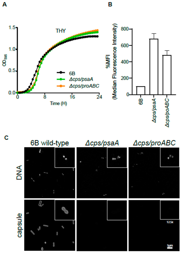
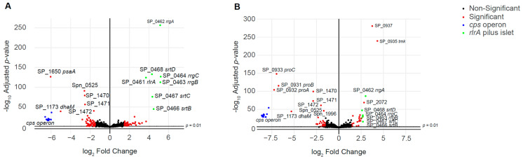
Methods: Strains containing cps locus deletions combined with the S. pneumoniae virulence factors psaA (reduces colonisation) or proABC (no effect on colonisation) were constructed and their virulence phenotypes and ability to prevent recolonisation or invasive infection assessed using mouse infection models. Serological responses to colonisation were compared between strains using ELISAs, immunoblots and 254 S. pneumoniae protein antigen array.

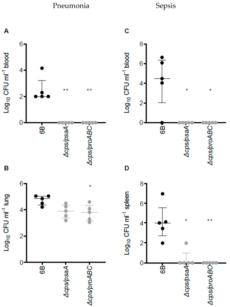
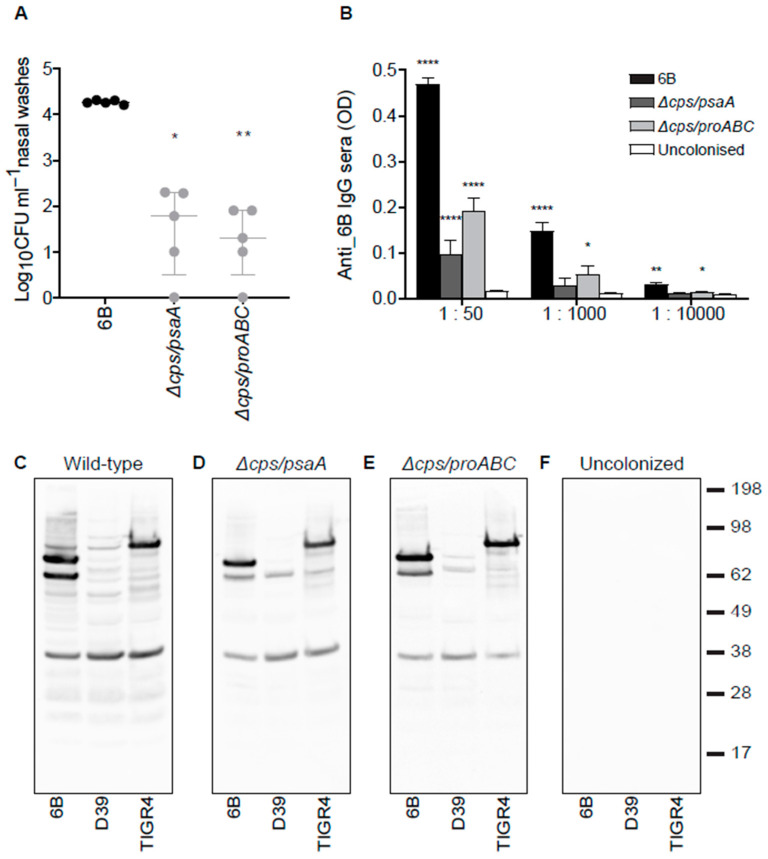
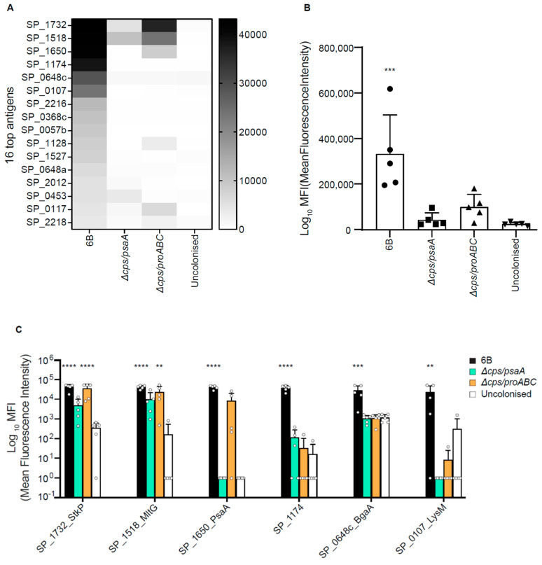
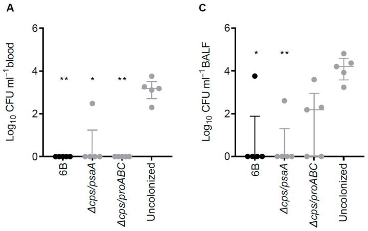
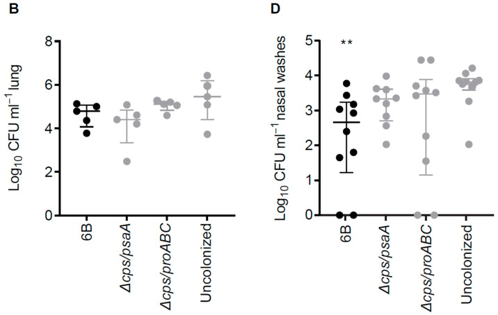
Measurements and main results: The ∆cps/piaA and ∆cps/proABC strains were strongly attenuated in virulence in both invasive infection models and had a reduced ability to colonise the nasopharynx. ELISAs, immunoblots and protein arrays showed colonisation with either strain stimulated weaker serological responses than the wild type strain. Mice previously colonised with these strains were protected against septicaemic pneumonia but, unlike mice colonised with the wild type strain, not against S. pneumoniae recolonisation.

Conclusions: Colonisation with the ∆cps/piaA and ∆cps/proABC strains prevented subsequent septicaemia, but in contrast, to published data for encapsulated double mutant strains they did not prevent recolonisation with S. pneumoniae. These data suggest targeting the cps locus is a less effective option for creating live attenuated strains that prevent S. pneumoniae infections.

Keywords: Streptococcus pneumoniae; capsule; colonisation; immunity; proABC; psaA; vaccine.


Conflict of interest statement

The authors declare no conflict of interest.

Figures

Figure 1
Figure 1
Phenotypic characterization of double unencapsulated mutants ∆cps/psaA and ∆cps/proABC. (A) Growth of wild type and unencapsulated strains ∆cps/psaA and ∆cps/proABC in THY assessed by measuring OD595 for a 24 h period. (B) Effect of the S. pneumoniae capsule on C3b deposition. Median fluorescence intensity (MFI) of C3b deposition measured using flow cytometry on the wild type 6B and unencapsulated mutants when incubated in 25% human serum. Error bars represent SDs and asterisks represent the differences between wild type and the unencapsulated mutant strain. For both mutant strains, p is < 0.0001 (one way ANOVA) compared to the wild type strain. (C) Fluorescent microscopy of wild type and unencapsulated double mutant strains following incubation with 4′,6-diamidino-2-phenylindole (DAPI) (binds to DNA to identify bacterial cells, top panels) or pneumococcal antiserum labelled with Alexa fluor 546 (recognizes serotype 6 capsule, bottom panels). The scale bar (bottom right) represents 5 µm and the inserts show a 12.5 higher magnification of selected bacteria.
Figure 2
Figure 2
Virulence of double unencapsulated mutant strains in murine pneumonia and sepsis models. (A,B) Pneumonia model; CFU obtained from blood (A) and lung (B) of CD1 mice 28 h post intranasal inoculation with 1 × 10⁷ CFU of wild type 6B or double mutant S. pneumoniae strains. (C) and (D) Sepsis model; CFU in blood (C) or spleen (D) of CD1 mice 24 h post intraperitoneal inoculation with 5 × 10⁶ CFU of wild type 6B or double mutant S. pneumoniae strains. Each symbol represents CFU data from a single mouse (black symbols, wild type S. pneumoniae infection, grey mutant S. pneumoniae strain infection), horizontal bars represent median values, error bars represent interquartile range and asterisks represent statistical significance compared to the wild type strain (Kruskal–Wallis with Dunn’s post hoc test to identify significant differences between groups, * p < 0.05; ** p < 0.01).
Figure 3
Figure 3
Volcano plots showing differential expression of genes in the live attenuated strains. Log2 ratio gene expression levels for the mutant strains (A) ∆cps/psaA and (B) ∆cps/proABC compared to the wild type strain. Fold change expression is represented in the X-axis, with negative values indicating genes that are under-expressed in the mutant versus the wild type, while positive fold change values indicate genes that are over-expressed in the mutant versus the wild type. The Y-axis shows the p values (as -log10 values) for differentially expressed genes. Genes belonging to cps locus are shown in blue, while those belonging to the pilus are shown in green, other genes that are differentially expressed are represented in red and all other genes are represented in black. Differential gene expression and statistical significance were analysed using the DESeq2 method. The dashed line above the x axis marks the significance threshold of p = 0.01. A full list of the genes showing differential expression with their expression levels is shown in Table S2, with selected genes of interest shown in Table 1.
Figure 4
Figure 4
Wild type 6B and the unencapsulated double mutant strains induce a systemic antibody response after nasopharynx colonisation. (A) Colonisation model; nasal wash CFU 7 days post colonisation of CD1 mice with 1 × 10⁷ CFU of wild type 6B or the double mutant S. pneumoniae strains. (B) Whole-cell enzyme-linked immunosorbent assay (ELISA) anti-6B immunoglobulin (Ig)G responses in mouse sera 28 days post-colonisation with the corresponding strain 6B (black bars), ∆cps/psaA mutant (dark grey bars), ∆cps/proABC mutant (light grey bars) compared with uncolonised controls (white bars). N = 5 for each group and the data analysed using Kruskal-Wallis with Dunn’s post hoc test to identify significant differences between selected groups; *, p < 0.05; **, p < 0.01 ****, p < 0.0001. (CF) IgG immunoblots for whole-cell lysates of three different S. pneumoniae strains (6B, D39 and TIGR4) probed with sera obtained 28 days after two episodes of colonisation with 6B (C), ∆csp/psaA (D), ∆cps/proABC (E) strains, or from PBS sham colonized mice (F).
Figure 5
Figure 5
Identification of the protein antigens recognized by IgG in serum from mice colonised with wild type and double mutant S. pneumoniae strains. Antigen specific IgG binding data obtained by probing a protein array containing 254 S. pneumoniae protein antigens with sera from mice colonised twice with the 6B, ∆cps/psaA, or ∆cps/proABC strains. (A) Heatmap of mean IgG binding levels to the top 16 proteins recognised by IgG in colonised mouse sera (n = 5 mice). (B) Aggregated MFI for IgG binding to the 30 antigens with the highest level of IgG binding in serum from mice colonised twice with the 6B, ∆cps/psaA, or ∆cps/proABC strains, or sham colonized with PBS. Each symbol represents data from 1 mouse, bars represent mean and error bars SD. Asterisks represent statistical significance compared to the uncolonised group (Kruskal-Wallis with Dunn’s multiple comparisons posthoc test; ***, p < 0.001). (C) Binding results in sera from mice colonised twice with the 6B strain, ∆cps/psaA, or ∆cps/proABC strains, or sham colonised to the 6 antigens with the highest level of IgG binding in serum obtained from wild type colonized mice. Each symbol represents data from 1 mouse, bars represent mean and error bars SD. Asterisks represent statistical significance compared to the uncolonised group (two-way ANOVA with Dunnett’s for multiple comparisons, **, p < 0.01; ***, p < 0.001, ****, p < 0.0001).
Figure 6
Figure 6
Rechallenge of mice previously colonised with 6B and the double mutant attenuated strains using wild type 6B and pneumonia and colonisation models. (AC) Target organ CFU for sham-colonised, 6B colonised, or double mutant strain colonised CD1 mice challenged 30 days after colonisation by intranasal inoculation of 1 × 10⁷ CFU wild type 6B S. pneumoniae. (A) Blood, (B) lung homogenate and (C) BALF S. pneumoniae CFU (log10 ml−1) 24 hours following the pneumonia challenge. (D) Nasal wash CFU 7 days after intranasal recolonisation challenge of CD1 mice with 1 × 10⁷ CFU of the S. pneumoniae 6B strain 42 days after two episodes of colonisation with the wild type 6B or double mutant strains. Each symbol represents data from a single mouse, horizontal bars represent median values, error bars represent interquartile range and asterisks represent statistical significance compared to sham colonised group (Kruskall–Wallis with Dunn’s post hoc test; *, p < 0.05; **, p < 0.01).
Figure 6
Figure 6
Rechallenge of mice previously colonised with 6B and the double mutant attenuated strains using wild type 6B and pneumonia and colonisation models. (AC) Target organ CFU for sham-colonised, 6B colonised, or double mutant strain colonised CD1 mice challenged 30 days after colonisation by intranasal inoculation of 1 × 10⁷ CFU wild type 6B S. pneumoniae. (A) Blood, (B) lung homogenate and (C) BALF S. pneumoniae CFU (log10 ml−1) 24 hours following the pneumonia challenge. (D) Nasal wash CFU 7 days after intranasal recolonisation challenge of CD1 mice with 1 × 10⁷ CFU of the S. pneumoniae 6B strain 42 days after two episodes of colonisation with the wild type 6B or double mutant strains. Each symbol represents data from a single mouse, horizontal bars represent median values, error bars represent interquartile range and asterisks represent statistical significance compared to sham colonised group (Kruskall–Wallis with Dunn’s post hoc test; *, p < 0.05; **, p < 0.01).

Similar articles

Cited by

References

    1. Lim W.S., Macfarlane J.T., Boswell T.C., Harrison T.G., Rose D., Leinonen M., Saikku P. Study of community acquired pneumonia aetiology (SCAPA) in adults admitted to hospital: Implications for management guidelines. Thorax. 2001;56:296–301. doi: 10.1136/thorax.56.4.296. - DOI - PMC - PubMed
    1. Melegaro A., Edmunds W.J., Pebody R., Miller E., George R. The current burden of pneumococcal disease in England and Wales. J. Infect. 2006;52:37–48. doi: 10.1016/j.jinf.2005.02.008. - DOI - PubMed
    1. Garcha D.S., Thurston S.J., Patel A.R.C., Mackay A.J., Goldring J.J.P., Donaldson G.C., McHugh T.D., Wedzicha J.A. Changes in prevalence and load of airway bacteria using quantitative PCR in stable and exacerbated COPD. Thorax. 2012;67:1075–1080. doi: 10.1136/thoraxjnl-2012-201924. - DOI - PubMed
    1. Moberley S., Holden J., Tatham D.P., Andrews R.M. Vaccines for preventing pneumococcal infection in adults. Cochrane Database Syst. Rev. 2013;2013:CD000422. doi: 10.1002/14651858.CD000422.pub3. - DOI - PMC - PubMed
    1. Bonten M.J., Huijts S.M., Bolkenbaas M., Webber C., Patterson S., Gault S., Van Werkhoven C.H., Van Deursen A.M., Sanders E.A., Verheij T.J., et al. Polysaccharide conjugate vaccine against pneumococcal pneumonia in adults. N. Engl. J. Med. 2015;372:1114–1125. doi: 10.1056/NEJMoa1408544. - DOI - PubMed