Case Reports
. 2021 Mar 15;10(3):343.
doi: 10.3390/pathogens10030343.

Malacoplakia of the Uterine Cervix: A Case Report

Affiliations
Case Reports

Malacoplakia of the Uterine Cervix: A Case Report

Adela Saco et al. Pathogens. .

Abstract

Malacoplakia is an uncommon chronic granulomatous inflammation that rarely affects the female genital tract. A case of a 78-year-old woman with malacoplakia involving the uterine cervix and the vagina is described. The patient complained of vaginal bleeding. Clinically, a 13-mm mass was detected in the cervix, which was confirmed by ultrasound scan and magnetic resonance imaging. Histological examination showed a dense histiocytic infiltrate with abundant Michaelis-Gutmann bodies involving the uterine cervix and the upper vagina. The presence of Escherichia coli was confirmed in the lesion by immunohistochemistry and polymerase chain reaction. Only 12 cases of cervical malacoplakia have been reported to date. This condition should be included in the differential diagnosis of cervical tumors.

Keywords: Michaelis–Gutmann bodies; inflammatory pathology; malacoplakia; uterine cervix.


Conflict of interest statement

The authors declare no conflict of interest.

Figures

Figure 1
Figure 1
Cervicovaginal Pap-smear with abundant histiocytes and neutrophils (Papanicolaou stain, 400×).
Figure 2
Figure 2
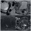
Pelvic magnetic resonance imaging (MRI). An infiltrative cervical mass (arrow) is seen in axial (A) and sagittal (B) T2 weighted MRI with stromal infiltration as a disruption of the cervical stroma hypointensity and focal infiltration of superior vaginal third (arrow in A). The lesion showed restricted diffusion in high b value (C: DWI b 800) and contrast enhancement (D: Post-contrast axial T1w fat saturated MRI).
Figure 3
Figure 3
Macroscopic image showing a yellowish lesion with poorly defined margins involving the cervix and upper vagina.
Figure 4
Figure 4
(A) Inflammatory infiltrate replacing the lamina propria and ulcerating the superficial epithelium (hematoxylin and eosin, 40×); (B) Histiocytic infiltrate in the cervical stroma (hematoxylin and eosin, 200×); (C) Immunohistochemical stain for CD68 showing a dense infiltrate composed by macrophages (immunohistochemical stain anti CD68); (D) Abundant Michaelis–Gutmann bodies (hematoxylin ad eosin 200×); (E) Presence of intra and extracytoplasmic Michaelis–Gutmann bodies positive for von Kossa stain (100×); (F) Escherichia coli bacilli present inside the cytoplasm of histiocytes (immunohistochemical stain anti E. coli, 600×).

References

    1. Michaelis L., Gutmann C. Ueber Einschlüsse in Blasentumoren. Ztschr. Klin. Med. 1902;47:208–215.
    1. Darvishian F., Teichberg S., Meyersfield S., Urmacher C.D. Concurrent malakoplakia and papillary urothelial carcinoma of the urinary bladder. Ann. Clin. Lab. Sci. 2001;31:147–150. - PubMed
    1. Lin J.I., Caracta P.F., Chang C.H., Uchwat F., Tseng C.H. Malacoplakia of the Vagina. South. Med. J. 1979;72:326–328. doi: 10.1097/00007611-197903000-00026. - DOI - PubMed
    1. Thomas W., Sadeghieh B., Fresco R., Rubenstone A.I., Stepto R.C., Carasso B. Malacoplakia of the Endometrium, a Probable Cause of Postmenopausal Bleeding. Am. J. Clin. Pathol. 1978;69:637–641. doi: 10.1093/ajcp/69.6.637. - DOI - PubMed
    1. Stewart C.J.R., Thomas M.A. Malacoplakia of the Uterine Cervix and Endometrium. Cytopathology. 1991;2:271–275. doi: 10.1111/j.1365-2303.1991.tb00498.x. - DOI - PubMed

Publication types