. 2021 Mar 20;22(6):3166.
doi: 10.3390/ijms22063166.

Development of a New Highly Selective Monoclonal Antibody against Preferentially Expressed Antigen in Melanoma (PRAME) and Identification of the Target Epitope by Bio-Layer Interferometry

Affiliations

Development of a New Highly Selective Monoclonal Antibody against Preferentially Expressed Antigen in Melanoma (PRAME) and Identification of the Target Epitope by Bio-Layer Interferometry

Jwala Priyadarsini Sivaccumar et al. Int J Mol Sci. .

Abstract

Background: Monoclonal antibodies (mAbs) against cancer biomarkers are key reagents in diagnosis and therapy. One such relevant biomarker is a preferentially expressed antigen in melanoma (PRAME) that is selectively expressed in many tumors. Knowing mAb's epitope is of utmost importance for understanding the potential activity and therapeutic prospective of the reagents.

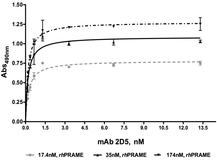
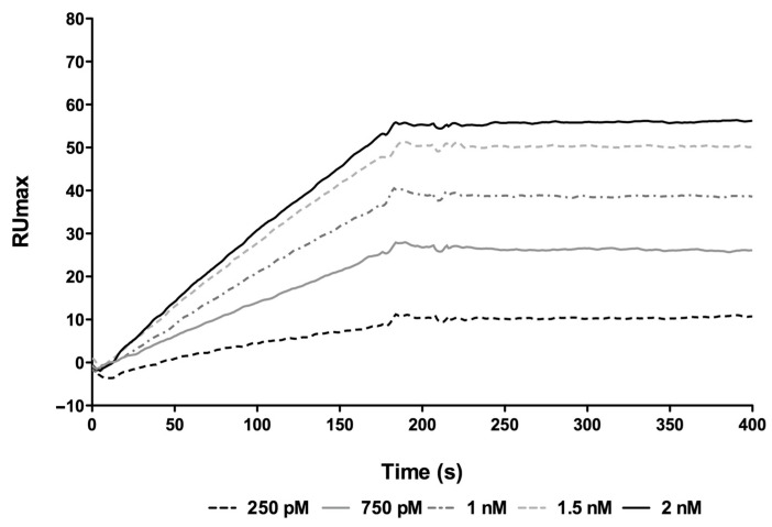
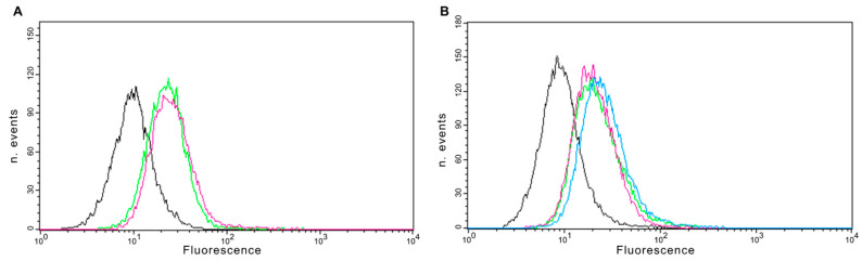
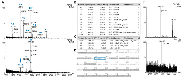
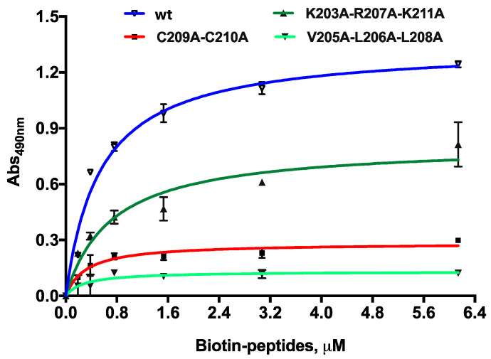
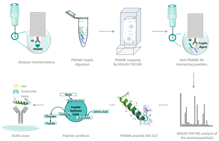
Methods: We generated a mAb against PRAME immunizing mice with PRAME fragment 161-415; the affinity of the antibody for the protein was evaluated by ELISA and SPR, and its ability to detect the protein in cells was probed by cytofluorimetry and Western blotting experiments. The antibody epitope was identified immobilizing the mAb on bio-layer interferometry (BLI) sensor chip, capturing protein fragments obtained following trypsin digestion and performing mass spectrometry analyses.

Results: A mAb against PRAME with an affinity of 35 pM was obtained and characterized. Its epitope on PRAME was localized on residues 202-212, taking advantage of the low volumes and lack of fluidics underlying the BLI settings.

Conclusions: The new anti-PRAME mAb recognizes the folded protein on the surface of cell membranes suggesting that the antibody's epitope is well exposed. BLI sensor chips can be used to identify antibody epitopes.

Keywords: PRAME; bio-layer interferometry; epitope identification; mAb.


Conflict of interest statement

The authors declare no conflict of interest.

Figures

Figure 1
Figure 1
ELISA testing of the binding between the anti-preferentially expressed antigen in melanoma (PRAME) 2D5 mAb and rhPRAME) coated at the reported concentrations. A KD of 0.19 ± 0.03 nM was roughly estimated as the average value of the least 3 independent experiments.
Figure 2
Figure 2
SPR dose–response assays between the immobilized rhPRAME and the anti-PRAME 2D5 mAb at various concentrations. The overlaid sensorgrams obtained at mAb concentrations between 250 pM and 2.0 nM are shown. All experiments were carried out at 25 °C at a constant flow rate of 20 μL/min using HBS-EP as a running buffer. Kinetic parameters are reported in Table 1.
Figure 3
Figure 3
(A) Western blotting analysis of endogenous PRAME in L1236 (PRAME-positive) and KG1 (PRAME-negative) cell extracts using the mAb 2D5 at various concentrations. (B) Western blotting analysis of U2OS cell extracts using the mAb 2D5 at 5.0 µg/mL. In (A) M: markers; 1: KG1 cell extract probed with 2D5 at 5.0 µg/mL; 2: L1236 cell extract probed with 2D5 at 5.0 µg/mL; 3: KG1 cell extract probed with 2D5 at 2.0 µg/mL. 4: L1236 cell extract probed with 2D5 at 2.0 µg/mL; 5: KG1 cell extract probed with 2D5 at 1.0 µg/mL; 6: L1236 cell extract probed with 2D5 at 1.0 µg/mL. In (B) M: markers; 1: U20S (PRAME-positive) cell extract; 2: rhPRAME used as a positive control.
Figure 4
Figure 4
Flow cytometry analysis of U2OS cells using the 2D5 monoclonal antibody and the commercial antibody code ab89097, used as a positive control. (A) Cells at a density of 5 × 10⁷ cells/mL were incubated with 15 µg/mL of the commercial anti-PRAME antibody (green curve) or 2D5 mAb (magenta curve). (B) Cells were incubated with 7.5 (magenta curve), 15 (green curve) or 30 µg/mL (blue curve) of anti-PRAME 2D5 mAb. Black curves represent the cells incubated with isotype control antibody. These pictures are representative of different independent experiments.
Figure 5
Figure 5
(A) Matrix-assisted laser desorption ionization time-of-flight (MALDI-TOF) spectra in the m/z range 1000–3000 of tryptic peptides (upper panel) and antibody-bound eluted peptides (lower panel) of human recombinant PRAME protein. (B) List of tryptic peptides from human recombinant PRAME protein (Pr) and human keratins (K) as identified by MALDI-TOF MS analyses on the digested protein. (C) List of tryptic peptides from the antibody-bound fraction eluted from the sensor tip as identified by MALDI-TOF MS analysis. Peptide’s theoretical and experimental masses, along with mass errors, amino acid positions and modification and the number of trypsin missed-cleavage sites, are reported (*, peptide ions not shown in MALDI-TOF spectra). (D) Tryptic peptides identified by MALDI-TOF MS analysis and mapping on the PRAME protein region 161–415. Sequence regions corresponding to the mapped tryptic peptides are shaded gray. The His-tag-containing linker (His-Tag) at the protein N-terminus is bold and underlined. The protein region 202–212, corresponding to the peptide uniquely detected in the antibody-bound sample, is blue boxed. (E) Magnification of the m/z region containing the ion signal at m/z 1446.92, uniquely detected in the sample of antibody-bound peptides.
Figure 6
Figure 6
Dose–response ELISA binding of the biotin-PRAME (202–212) wild-type and mutanted peptides to the coated 2D5 mAb. For the wild type peptide, an affinity constant of 0.55 ± 0.10 μM was roughly estimated. The binding to the mutated variants under the same conditions are also reported.
Scheme 1
Scheme 1
Schematic representation of the approach followed to identify the epitope of mAb 2D5.

Similar articles

Cited by

References

    1. Sivaccumar J., Sandomenico A., Vitagliano L., Ruvo M. Monoclonal antibodies: A prospective and retrospective view. Curr. Med. Chem. 2021;28:435–471. doi: 10.2174/0929867327666200219142231. - DOI - PubMed
    1. Ikeda H., Lethe B., Lehmann F., van Baren N., Baurain J.F., de Smet C., Chambost H., Vitale M., Moretta A., Boon T., et al. Characterization of an antigen that is recognized on a melanoma showing partial HLA loss by CTL expressing an NK inhibitory receptor. Immunity. 1997;6:199–208. doi: 10.1016/S1074-7613(00)80426-4. - DOI - PubMed
    1. Al-Khadairi G., Decock J. Cancer Testis Antigens and Immunotherapy: Where Do We Stand in the Targeting of PRAME? Cancers. 2019;11:984. doi: 10.3390/cancers11070984. - DOI - PMC - PubMed
    1. Abdelmalak C.A., Yahya R.S., Elghannam D.M., El-Khadragy A.E., Abd El Messih H.M. PRAME gene expression in childhood acute lymphoblastic leukemia: Impact on prognosis. Clin. Lab. 2014;60:55–61. doi: 10.7754/Clin.Lab.2013.121137. - DOI - PubMed
    1. Zhang Y.H., Lu A.D., Yang L., Li L.D., Chen W.M., Long L.Y., Zhang L.P., Qin Y.Z. PRAME overexpression predicted good outcome in pediatric B-cell acute lymphoblastic leukemia patients receiving chemotherapy. Leuk. Res. 2017;52:43–49. doi: 10.1016/j.leukres.2016.11.005. - DOI - PubMed

MeSH terms

LinkOut - more resources