Review
. 2021 Mar 20;10(3):687.
doi: 10.3390/cells10030687.

Cell-Matrix Interactions in the Eye: From Cornea to Choroid

Affiliations
Review

Cell-Matrix Interactions in the Eye: From Cornea to Choroid

Andrew E Pouw et al. Cells. .

Abstract

The extracellular matrix (ECM) plays a crucial role in all parts of the eye, from maintaining clarity and hydration of the cornea and vitreous to regulating angiogenesis, intraocular pressure maintenance, and vascular signaling. This review focuses on the interactions of the ECM for homeostasis of normal physiologic functions of the cornea, vitreous, retina, retinal pigment epithelium, Bruch's membrane, and choroid as well as trabecular meshwork, optic nerve, conjunctiva and tenon's layer as it relates to glaucoma. A variety of pathways and key factors related to ECM in the eye are discussed, including but not limited to those related to transforming growth factor-β, vascular endothelial growth factor, basic-fibroblastic growth factor, connective tissue growth factor, matrix metalloproteinases (including MMP-2 and MMP-9, and MMP-14), collagen IV, fibronectin, elastin, canonical signaling, integrins, and endothelial morphogenesis consistent of cellular activation-tubulogenesis and cellular differentiation-stabilization. Alterations contributing to disease states such as wound healing, diabetes-related complications, Fuchs endothelial corneal dystrophy, angiogenesis, fibrosis, age-related macular degeneration, retinal detachment, and posteriorly inserted vitreous base are also reviewed.

Keywords: AMD; MMP-14; MMP-9; TGF-beta; TIMP-3; VEGF; bruch’s membrane; choroid; collagen; descemet membrane; interphotoreceptor matrix; metalloproteinases.


Conflict of interest statement

The authors declare no conflict of interest related to this work. E.H.S. has received research funding from Oxford BioMedica. The funders had no role in the design of the study; in the collection, analyses, or interpretation of data; in the writing of the manuscript, or in the decision to publish the results.

Figures
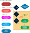
Figure 1
Figure 1
Corneal stromal wound healing. Stromal wound healing in the cornea is mediated by signaling of transforming growth factor-beta (TGF-β), matrix metalloproteinases (MMPs), and a balance between pro-angiogenic and anti-angiogenic factors. In some cases, corneal neovascularization can occur.
Figure 2
Figure 2
Corneal neovascularization with stromal scarring secondary to atopic keratoconjunctivitis.
Figure 3
Figure 3
Extracellular fibrosis (arrowhead) and dense guttae (**) in the posterior cornea secondary to Fuchs endothelial corneal dystrophy.
Figure 4
Figure 4
Configuration of the trabecular meshwork. In both the corneoscleral and uveoscleral layers, the trabecular meshwork cells wrap around a core of extracellular matrix components. Note the increasingly large intertrabecular pores between the trabecular meshwork beams in the deeper layers. In the juxtacanalicular layer, the extracellular matrix and the trabecular meshwork cells have a more irregular and interwoven spatial relationship.
Figure 5
Figure 5
Overview of cellular and extracellular matrix interactions in glaucomatous tissue remodeling. The interplay of numerous factors, including environmental stressors, enzymatic reactions, growth factors, glycoproteins and proteoglycans as well as cytoskeletal elements all contribute to a feedback loop where outflow facility is disturbed. Red circles indicate negative situational change. Green circle indicates positive situational change. Dark blue diamonds highlight critical signaling factors. Light blue diamonds indicate extracellular matrix glycoproteins and proteoglycans. Red rhombuses indicate negative catalysts. Green rhombus indicates positive catalysts. White circle indicates exogenous factors. ECM: Extracellular matrix; TGF: Transforming growth factor; CTGF: Connective tissue growth factor; α-SMA: Alpha smooth muscle actin; CLANs: Cross-linked actin networks; TIMP-1: Tissue inhibitors of metalloproteinases 1.
Figure 6
Figure 6
Schematic of vitreous base inserting into the retina. At the pars plana normally (A), when it is posteriorly inserted (B), or at posterior the equator, averaging 7.6 mm posterior to the ora serrata which predisposes to more retinal tears (C). Adapted from Sohn et al. [111].
Figure 7
Figure 7
Insoluble interphotoreceptor matrix glycoproteins, the gene products for IMPG1 and IMPG2 are distributed in domains surrounding rod and cone photoreceptors. The relative distributions of cone matrix sheaths labeled with peanut agglutinin (red) is depicted compared to rod outer segments labeled with anti-rhodopsin (green).
Figure 8
Figure 8
Extracellular matrix mediated endothelial morphogenesis. The diagram illustrates the concept of “fire and ice” representing balance of extracellular matrix-based signaling which dictates endothelial cellular activation and tubulogenesis with endothelial cell stabilization. The complex process is mediated by the interaction between extracellular matrix components (including collagen I, fibrin, fibronectin and laminin) and various integrins. Abbreviations: VEGF: vascular endothelial growth factor; TGF-β: transforming growth factor beta.
Figure 9
Figure 9
Representative hematoxylin and eosin (H&E) and immunofluorescence images from four patients’ membranes in a randomized controlled trial. Co-labeling of antibodies for (A) CD31 (Green)-CTGF (Red) and (B) cytokeratin (Green)-VEGF (Red). Note the H&E-stained sections do not correspond precisely to the cytokeratin-labeled sections. While intravitreal bevacizumab did not significantly decrease CTGF (A-top panels) or VEGF (B-top panels) expression in membranes compared to sham group, VEGF was still expressed in membranes of eyes given bevacizumab (B, right panels). Scale bar = 100 μm. Abbreviations: CTGF: connective Tissue Growth Factor; VEGF: vascular endothelial growth factor. Adapted from Jiao et al. [174].
Figure 10
Figure 10
Anti-collagen IV labeling in human donor eyes. Drusen (asterisks) associated with aging do not show labeling with antibodies directed against collagen type IV (AC). Laminae within the autosomal dominant radial drusen are immunoreactive with anti-collagen IV antibodies (green fluorescence). Sections were also labeled with DAPI (blue nuclear fluorescence) and were exposed in the rhodamine channel (red autofluorescence of the RPE). Scalebar = 50 μm. Adapted from Sohn et al. [183].
Figure 11
Figure 11
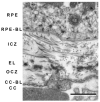
Transmission electron micrograph depicting the layers of Bruch’s membrane from a human eye. Both the basal laminae of the RPE and choriocapillaris (RPE-BL and CC-BL) are depicted, in addition to inner collagenous zone (ICZ) and outer collagenous zone (OCZ), occupied by fibrillar collagens, as well as the elastic lamina (EL), evident by its thick electron dense bundles. Scale bar = 1 μm.

References

    1. Reinhard J., Joachim S.C., Faissner A. Extracellular matrix remodeling during retinal development. Exp. Eye Res. 2015;133:132–140. doi: 10.1016/j.exer.2014.07.001. - DOI - PubMed
    1. Yue B. Biology of the extracellular matrix: An overview. J. Glaucoma. 2014;23:S20–S23. doi: 10.1097/IJG.0000000000000108. - DOI - PMC - PubMed
    1. Hubmacher D., Apte S.S. The biology of the extracellular matrix: Novel insights. Curr. Opin. Rheumatol. 2013;25:65–70. doi: 10.1097/BOR.0b013e32835b137b. - DOI - PMC - PubMed
    1. Al-Ubaidi M.R., Naash M.I., Conley S.M. A perspective on the role of the extracellular matrix in progressive retinal degenerative disorders. Investig. Ophthalmol. Vis. Sci. 2013;54:8119–8124. doi: 10.1167/iovs.13-13536. - DOI - PMC - PubMed
    1. Chang J.H., Huang Y.H., Cunningham C.M., Han K.Y., Chang M., Seiki M., Zhou Z., Azar D.T. Matrix metalloproteinase 14 modulates signal transduction and angiogenesis in the cornea. Surv. Ophthalmol. 2016;61:478–497. doi: 10.1016/j.survophthal.2015.11.006. - DOI - PMC - PubMed

Publication types

MeSH terms

LinkOut - more resources