. 2021 Mar 20;10(3):690.
doi: 10.3390/cells10030690.

P-Glycoprotein Inhibitor Tariquidar Plays an Important Regulatory Role in Pigmentation in Larval Zebrafish

Affiliations

P-Glycoprotein Inhibitor Tariquidar Plays an Important Regulatory Role in Pigmentation in Larval Zebrafish

Natalia Kasica et al. Cells. .

Abstract

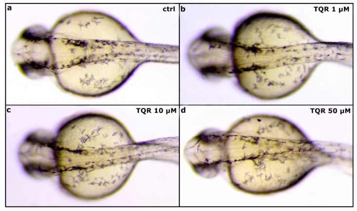
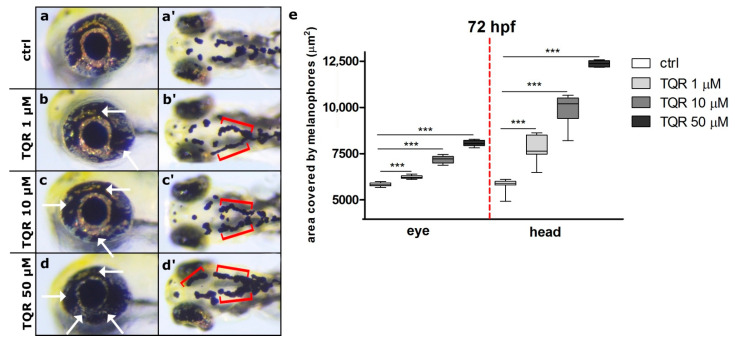
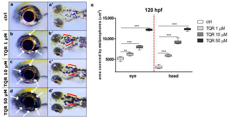
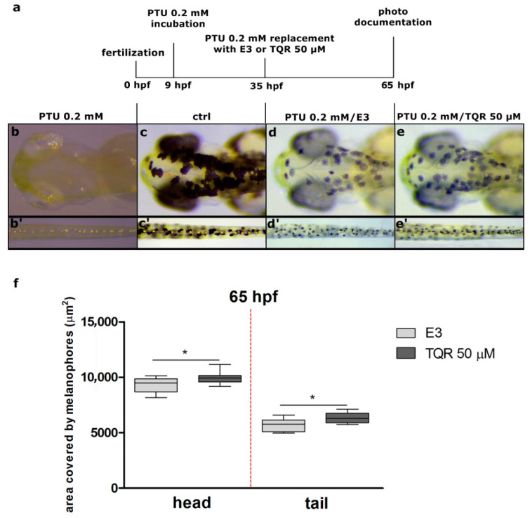
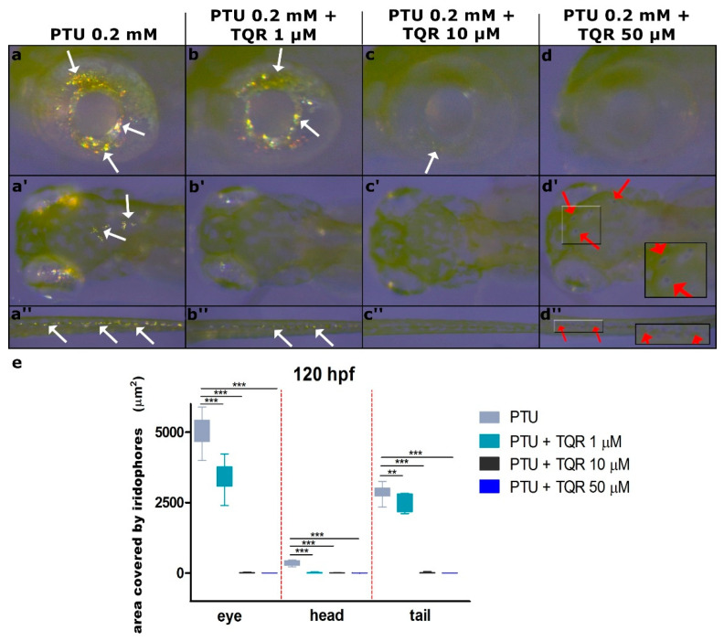
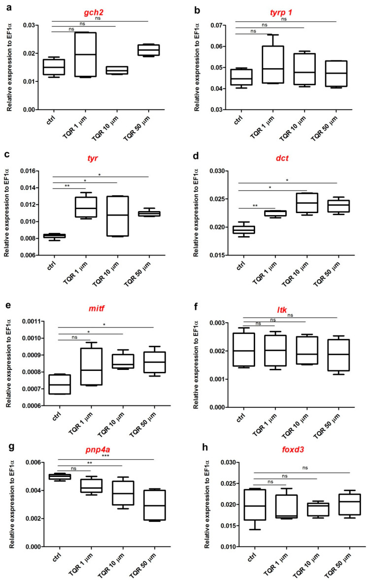
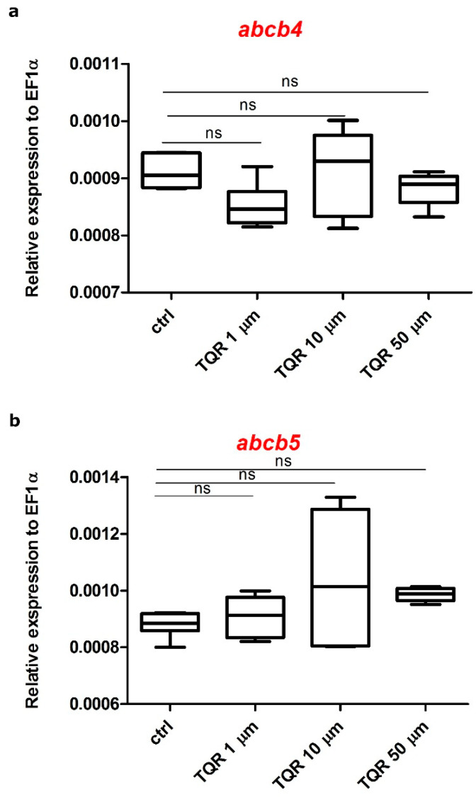
Zebrafish has emerged as a powerful model in studies dealing with pigment development and pathobiology of pigment diseases. Due to its conserved pigment pattern with established genetic background, the zebrafish is used for screening of active compounds influencing melanophore, iridophore, and xanthophore development and differentiation. In our study, zebrafish embryos and larvae were used to investigate the influence of third-generation noncompetitive P-glycoprotein inhibitor, tariquidar (TQR), on pigmentation, including phenotype effects and changes in gene expression of chosen chromatophore differentiation markers. Five-day exposure to increasing TQR concentrations (1 µM, 10 µM, and 50 µM) resulted in a dose-dependent augmentation of the area covered with melanophores but a reduction in the area covered by iridophores. The observations were performed in three distinct regions-the eye, dorsal head, and tail. Moreover, TQR enhanced melanophore renewal after depigmentation caused by 0.2 mM 1-phenyl-2-thiourea (PTU) treatment. qPCR analysis performed in 56-h post-fertilization (hpf) embryos demonstrated differential expression patterns of genes related to pigment development and differentiation. The most substantial findings include those indicating that TQR had no significant influence on leukocyte tyrosine kinase, GTP cyclohydrolase 2, tyrosinase-related protein 1, and forkhead box D3, however, markedly upregulated tyrosinase, dopachrome tautomerase and melanocyte inducing transcription factor, and downregulated purine nucleoside phosphorylase 4a. The present study suggests that TQR is an agent with multidirectional properties toward pigment cell formation and distribution in the zebrafish larvae and therefore points to the involvement of P-glycoprotein in this process.

Keywords: P-glycoprotein inhibitor; dopachrome tautomerase; iridophores; melanocyte inducing transcription factor; melanophores; pigment cells; purine nucleoside phosphorylase 4a; tariquidar; tyrosinase; zebrafish.


Conflict of interest statement

The authors declare that they have no competing interests.

Figures

Figure 1
Figure 1
Zebrafish melanophore pigmentation following the exposure to tariquidar (TQR) at 30 h post fertilization (hpf). A set of photographs presenting a dorsal view of 30 hpf zebrafish embryo displaying effects of tariquidar (TQR) exposure on the melanophore pigmentation within the dorsal head in four experimental groups: (a) control, (b) exposed to TQR 1 µM, (c) exposed to TQR 10 µM, and (d) exposed to TQR 50 µM. The developing dark pigment pattern does not show visible differences between the groups.
Figure 2
Figure 2
Dose-dependent increase in zebrafish melanophore pigmentation following the exposure to tariquidar (TQR) at 72 h post fertilization (hpf). A set of photographs presenting lateral and dorsal views of 72 hpf zebrafish larvae exhibiting effects of 68-h TQR exposure on the melanophore pigmentation within the eye and dorsal head in four experimental groups: (a,a’) control, (b,b’) exposed to TQR 1 µM (c,c’) exposed to TQR 10 µM, and (d,d’) exposed to TQR 50 µM. Control larvae presented normal melanophore distribution within both the eye and dorsal head (a,a’). Progressive hypermelanogenesis was observed as the concentration increased (b,b’,c,c’,d,d’). In the area of the eye, the melanophores started to occupy the area normally covered by the iridophores (b,c,d) (white arrows). Within the dorsal head in the control group, the melanophores were separated, while in the TQR exposed groups, they began to merge and form closely apposed groups (b’,c’,d’) (red buckle). (e) A graph presenting the area covered by melanophores (µm2) measured at 72 h post fertilization (hpf) within the eye and dorsal head (one-way ANOVA/Kruskal–Wallis, GraphPad Prism 5, *** p < 0.001).
Figure 3
Figure 3
Dose-dependent increase in zebrafish melanophore pigmentation following exposure to tariquidar (TQR) at 120 h post fertilization (hpf). A set of photographs presenting lateral and dorsal views of 120 hpf zebrafish larvae displaying effects of 116 h TQR exposure on the melanophore pigmentation within the eye and dorsal head in four experimental groups: (a,a’) control, (b,b’) exposed to TQR 1 µM, (c,c’) exposed to TQR 10 µM, and (d,d’) exposed to TQR 50 µM. The control larvae presented normal melanophore distribution within both the eye and dorsal head (a,a’). At 120 hpf, the effects of TQR exposure were the continuation of those observed at 72 hpf; however, they were more intense. Progressive hypermelanogenesis was observed with increasing concentration (b,b’;c,c’;d,d’). In the area of the eye, the melanophores began to occupy the area normally covered by the iridophores (b,c,d) (white arrows). Within the dorsal head in the control group, the melanophores were separated and small, while in the TQR exposed groups they were expanded and started to merge creating form closely apposed groups (b’,c’,d’) (red buckle). Additionally, in the control (a’) and 1 µM TQR-exposed group (a’) in the central part of the head, the iridophores were visible (blue arrows), while in 10 µM TQR- and 50 µM TQR-exposed groups these cells were not found. (e) A graph presenting the area covered by melanophores (µm2) measured at 120 h post fertilization (hpf) within the eye and dorsal head (one-way ANOVA/Kruskal–Wallis, GraphPad Prism 5, *** p < 0.001, ** p < 0.01).
Figure 4
Figure 4
A set of pictures taken at 65 h post fertilization (hpf) showing the melanogenic stimulatory effect of 50 µM tariquidar (TQR) after 0.2 mM 1-phenyl-2-thiourea (PTU) exposure in larval zebrafish. (a) A schematic representation of the schedule of pigmentation rescue investigation. Nine-hpf embryos were pretreated with 0.2 mM PTU. At 35 hpf, all the embryos were washed and immersed in the E3 medium or 50 µM TQR solution. The incubation after medium replacement lasted until 60 hpf. Photographs present (b,b’) larvae treated with PTU from 9 to 65 hpf, (c,c’) control larvae incubated in E3, (d,d’) larvae treated with PTU from 9 to 35 hpf and then replaced with E3, and (e,e’) larvae treated with PTU from 9 to 35 hpf and then replaced with TQR. (f) A graph presenting the area covered by melanophores (µm2) measured at 65 h post fertilization (hpf) within the dorsal head and tail. Replacement with 50 µM TQR resulted in a significant increase in the area covered by melanophores (µm2), within both the dorsal head and tail, in the TQR-exposed group compared to that determined in the E3 medium treated group (student’s t-test, GraphPad Prism 5, * p < 0.05).
Figure 5
Figure 5
Dose-dependent decrease in zebrafish iridophore pigmentation following the exposure to the mixture of 0.2 mM 1-phenyl-2-thiourea (PTU) and increasing concentrations of tariquidar (TQR) at 120 h post fertilization (hpf). A set of photographs showing lateral and dorsal views of 120 hpf zebrafish larvae exhibiting effects of 68 h PTU + TQR exposure on the iridophore pigmentation within the eye, dorsal head, and tail in four experimental groups: (a,a’,a”) exposed to PTU 0.2 mM, (b,b’,b”) exposed to PTU 0.2 mM + TQR 1 µM (c,c’,c”) exposed to PTU 0.2 mM + TQR 10 µM, and (d,d’,d”) exposed to PTU 0.2 mM + TQR 50 µM. PTU treatment resulted in melanophore disappearance with no influence on the iridophores (a,a’,a”; white arrows). Co-treatment with 1 µM TQR caused a slight reduction in the area covered with the iridophores; however, they were still well visible, mostly within the eye and tail (b,b”) (white arrows). In the 10-µM TQR co-treated group, only single iridophores were visible within the eye (c) (white arrow), while in the area of the dorsal head and tail they disappeared entirely (c’,c”). The addition of 50 µM TQR to the PTU solution resulted in a complete depletion of the iridophores within all areas investigated (d,d’,d”); however, small and faint melanophores were detectable (red arrows) (d’,d”). (e) A graph presenting the area covered by iridophores (µm2) measured at 120 h post fertilization (hpf) within the eye, dorsal head, and tail (Kruskall–Wallis, GraphPad Prism 5, *** p < 0.001, ** p < 0.01).
Figure 6
Figure 6
Expression profiles of chromatophore differentiation genes. The graphs show pooled data of the mRNA expression of (a) GTP cyclohydrolase 2 (gch2), (b) tyrosinase-related protein 1 (tyrp1), (c) tyrosinase (tyr), (d) dopachrome tautomerase (dct), (e) melanocyte inducing transcription factor (mitf), (f) leukocyte tyrosinase kinase (ltk), (g) purine nucleoside phosphorylase 4a (pnp4a), and (h) forkhead box D3(foxd3) from pooled 56-hour post-fertilization (hpf) wild-type zebrafish larvae (n = 30) in four experimental groups: 1) control, 2) exposed to TQR 1 µM, 3) exposed to TQR 10 µM, and 4) exposed to TQR 50 µM. Each group was covered by samples analyzed in triplicate in three separate experiments. Data in a figure represent the average of the three individual experiments. Gene expression values were normalized to housekeeping gene elongation factor 1-alpha (ef1-α). TQR had no statistically significant influence on gch2 (a), tyrp 1 (b), ltk (f), and foxd3 (h). Tyr (c) and dct (d) were significantly upregulated after the exposure to all studied TQR concentrations. Mitf (e) was not altered by TQR 1 µM; however, TQR in doses of 10 µM and 50 µM significantly increased the mRNA expression level. The pnp4a gene (g) was downregulated after the treatment with TQR, in dose-dependent manner (Kruskal–Wallis, GraphPad Prism 5, *** p < 0.001, ** p < 0.01, * p < 0.05, ns: not statistically significant differences (p > 0.05).
Figure 7
Figure 7
Expression profiles of zebrafish ATP-binding cassette (ABC) transporter family genes. The graphs show pooled data of the mRNA expression of (a) abcb4 and (b) abcb5 from pooled 56-hour post-fertilization (hpf) wild-type zebrafish larvae (n = 30) in four experimental groups: (1) control, (2) exposed to TQR 1 µM, (3) exposed to TQR 10 µM, and (4) exposed to TQR 50 µM. Each group was covered by samples analyzed in triplicate in three separate experiments. Data in the figure represent the average of the three individual experiments. Gene expression values were normalized to housekeeping gene elongation factor 1-alpha (Ef1-α). TQR had no statistically significant influence on both abcb4 (a) and abcb5 (b) (Kruskal–Wallis, GraphPad Prism 5, ns: not statistically significant differences (p > 0.05)).

References

    1. Cooper C.D. Insights from zebrafish on human pigment cell disease and treatment. Dev. Dyn. 2017;246:889–896. doi: 10.1002/dvdy.24550. - DOI - PubMed
    1. Pickart M.A., Sivasubbu S., Nielsen A.L., Shriram S., King R.A., Ekker S.C. Functional Genomics Tools for the Analysis of Zebrafish Pigment. Pigment. Cell Res. 2004;17:461–470. doi: 10.1111/j.1600-0749.2004.00189.x. - DOI - PubMed
    1. Rawls J.F., Mellgren E.M., Johnson S.L. How the Zebrafish Gets Its Stripes. Dev. Biol. 2001;240:301–314. doi: 10.1006/dbio.2001.0418. - DOI - PubMed
    1. Raible D.W., Wood A., Hodsdon W., Henion P.D., Weston J.A., Eisen J.S. Segregation and early dispersal of neural crest cells in the embryonic zebrafish. Dev. Dyn. 1992;195:29–42. doi: 10.1002/aja.1001950104. - DOI - PubMed
    1. Hirata M., Nakamura K.-I., Kondo S. Pigment cell distributions in different tissues of the zebrafish, with special reference to the striped pigment pattern. Dev. Dyn. 2005;234:293–300. doi: 10.1002/dvdy.20513. - DOI - PubMed

Publication types

MeSH terms

LinkOut - more resources