. 2021 Mar 20;11(3):221.
doi: 10.3390/jpm11030221.

Genetic Correction of IL-10RB Deficiency Reconstitutes Anti-Inflammatory Regulation in iPSC-Derived Macrophages

Affiliations

Genetic Correction of IL-10RB Deficiency Reconstitutes Anti-Inflammatory Regulation in iPSC-Derived Macrophages

Dirk Hoffmann et al. J Pers Med. .

Abstract

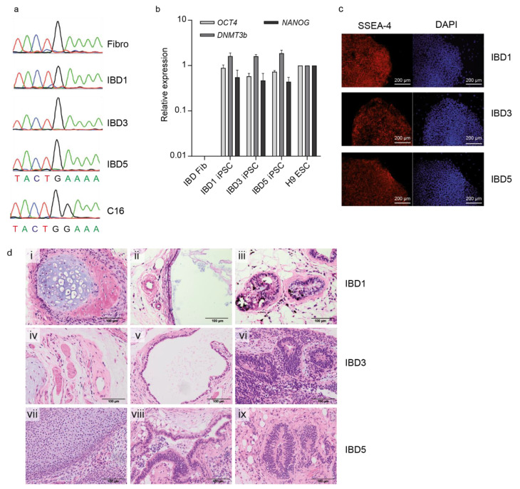
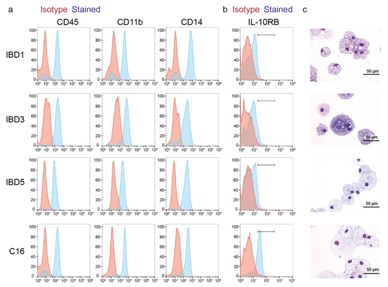
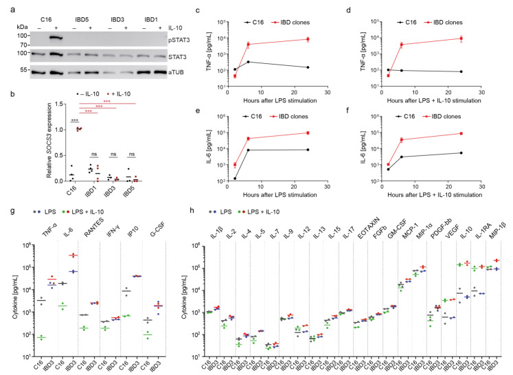
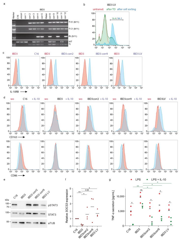
Patient material from rare diseases such as very early-onset inflammatory bowel disease (VEO-IBD) is often limited. The use of patient-derived induced pluripotent stem cells (iPSCs) for disease modeling is a promising approach to investigate disease pathomechanisms and therapeutic strategies. We successfully developed VEO-IBD patient-derived iPSC lines harboring a mutation in the IL-10 receptor β-chain (IL-10RB) associated with defective IL-10 signaling. To characterize the disease phenotype, healthy control and VEO-IBD iPSCs were differentiated into macrophages. IL-10 stimulation induced characteristic signal transducer and activator of transcription 3 (STAT3) and suppressor of cytokine signaling 3 (SOCS3) downstream signaling and anti-inflammatory regulation of lipopolysaccharide (LPS)-mediated cytokine secretion in healthy control iPSC-derived macrophages. In contrast, IL-10 stimulation of macrophages derived from patient iPSCs did not result in STAT3 phosphorylation and subsequent SOCS3 expression, recapitulating the phenotype of cells from patients with IL-10RB deficiency. In line with this, LPS-induced cytokine secretion (e.g., IL-6 and tumor necrosis factor-α (TNF-α)) could not be downregulated by exogenous IL-10 stimulation in VEO-IBD iPSC-derived macrophages. Correction of the IL-10RB defect via lentiviral gene therapy or genome editing in the adeno-associated virus integration site 1 (AAVS1) safe harbor locus led to reconstitution of the anti-inflammatory response. Corrected cells showed IL-10RB expression, IL-10-inducible phosphorylation of STAT3, and subsequent SOCS3 expression. Furthermore, LPS-mediated TNF-α secretion could be modulated by IL-10 stimulation in gene-edited VEO-IBD iPSC-derived macrophages. Our established disease models provide the opportunity to identify and validate new curative molecular therapies and to investigate phenotypes and consequences of additional individual IL-10 signaling pathway-dependent VEO-IBD mutations.

Keywords: IL-10 signaling; disease modeling; gene editing; gene therapy; very early-onset inflammatory bowel disease.


Conflict of interest statement

The authors declare no conflict of interest.

Figures

Figure 1
Figure 1
Generation of iPSCs from an IL-10RB-/- VEO-IBD patient. (a) Sanger sequencing to detect the IL-10RB mutation (c.G477A) in patient’s fibroblasts (Fib) and VEO-IBD iPSC clones (IBD1, IBD3, IBD5). Healthy C16 iPSC served as a control. (b) RT-qPCR expression analysis of endogenous pluripotency genes OCT4, NANOG, and DNMT3b in VEO-IBD iPSC and fibroblasts in relation to H9 ESCs (set to one, mean of technical replicates). (c) Immunofluorescence microscopy to detect pluripotency marker SSEA-4 expression on the surface of IBD iPSCs. DAPI was used for nuclei staining. Bars correspond to 200 µm. (d) Microscopic analyses of teratoma tissue generated from VEO-IBD iPSC clones after subcutaneous injection into immunodeficient mice. Hematoxylin and Eosin staining indicated mature tissues derived from all three germ layers: IBD1 (i)–(iii): cartilage (mesoderm, (i)), intestinal epithelium (endoderm, (ii)), neural tissue (ectoderm, (iii)). IBD3 (iv)–(vi): smooth muscle (mesoderm/ectoderm, (iv), ciliated airway epithelium (endoderm, (v)), neural tissue (ectoderm, (vi)). IBD5 (vii)–(ix): cartilage (mesoderm, (vii)), intestinal epithelium (endoderm, (viii)), neural tissue (ectoderm, (ix)). Bars correspond to 100 µm.
Figure 2
Figure 2
Macrophage differentiation of IL-10RB-/- VEO-IBD iPSCs. (a) Detection of macrophage surface marker expression (CD45, CD11b, CD14) on differentiated hematopoietic cells derived from healthy control (C16) and VEO-IBD (IBD1, IBD3, IBD5) iPSCs (red line, isotype; blue line, surface marker). (b) IL-10RB surface expression on iPSC-derived monocytes/macrophages analyzed by flow cytometry (red line, isotype; blue line, surface marker). (c) Morphological analyses by Pappenheim staining and light microscopy (bars correspond to 50 µm).
Figure 3
Figure 3
Deficient immune regulation in VEO-IBD iPSC-derived macrophages. (a) Detection of STAT3 phosphorylation (pY705; pSTAT3) upon LPS stimulation in iPSC-derived macrophages. Expression of STAT3 and alpha-Tubulin (aTUB) as controls. (b) Detection of IL-10-induced SOCS3 mRNA expression in healthy and IBD-iPSC-derived macrophages by qRT-PCR (mean of biological replicates, n = 3; *** p ≤ 0.001 two way-ANOVA and Holm–Šídák multiple comparison test). (c) Screening of TNF-α secretion by LPS-stimulated iPSC-derived macrophages over time using Bio-Plex assay (mean ± SD of IBD clones; n = 3). (d) Secretion of TNF-α upon LPS and IL-10 co-stimulation of macrophages over time (mean ± SD of IBD clones; n = 3). (e,f) Detection of IL-6 secretion by LPS, or LPS and IL-10 stimulated iPSC-derived macrophages (mean ± SD of IBD clones; n = 3). (g) Bio-Plex-based detection of IL-10 regulated inflammatory cytokines 6 h after LPS, or LPS and IL-10 co-stimulation of iPSC derived macrophages (biological replicates, n = 2). (h) Moderate or no effect of IL-10 on LPS-stimulated cytokine secretion as detected by Bio-Plex analysis (biological replicates, n = 2).
Figure 4
Figure 4
Genetic correction of iPSCs resulted in functional anti-inflammatory recovery of iPSC-derived macrophages. (a) Genotyping of the donor cassette integration into the AAVS1 genomic locus. PCR analysis using oligonucleotides to detect the right and left flanking arms of the donor cassette in IBD3 subclones to investigate 5′ and 3′ targeted integration (TI). Unspecific random integration (RI) and detection of the physiological wild type (WT) or targeted AAVS1 (TI) locus were used for more detailed validation of the loci. (b) Flow cytometric analysis of IL-10RB surface expression on lentiviral vector transduced IBD3 iPSC (IBD3 LV) before and after purification of transgene expressing cells (red line, untransduced iPSCs; green line, iPSCs after transduction (TD); blue line, sorted iPSCs by FACS). (c) Flow cytometric analysis of IL-10RB expression on corrected and uncorrected myeloid-differentiated suspension cells (red line, IBD3-derived cells; blue line, genetic corrected iPSC-derived cells). (d) IL-10 specific CD163 and CD86 up- or downregulation on uncorrected or corrected iPSC-derived macrophages upon stimulation (red line, without stimulation; blue line, stimulation with IL-10). (e) Western blot analysis of pSTAT3 phosphorylation (pY705, pSTAT3) in iPSC-derived macrophages after IL-10 stimulation. Expression of STAT3 and alpha-Tubulin (aTUB) as controls. (f) Upregulation of SOCS3 mRNA expression upon IL-10 stimulation in iPSC-derived macrophages detected by qRT-PCR (mean of biological replicates, n = 3; * p ≤ 0.05, ** p ≤ 0.01 unpaired t-test). (g) IL-10-mediated regulation of TNF-α secretion in LPS-stimulated uncorrected and corrected iPSC-derived macrophages (mean of biological replicates, n = 3; * p ≤ 0.05, ** p ≤ 0.01 two-way ANOVA and Holm–Šídák multiple comparison test).

Similar articles

Cited by

References

    1. Engelhardt K.R., Shah N., Faizura-Yeop I., Kocacik Uygun D.F., Frede N., Muise A.M., Shteyer E., Filiz S., Chee R., Elawad M., et al. Clinical outcome in IL-10– and IL-10 receptor–deficient patients with or without hematopoietic stem cell transplantation. J. Allergy Clin. Immunol. 2013;131:825–830.e9. doi: 10.1016/j.jaci.2012.09.025. - DOI - PubMed
    1. Glocker E.-O., Kotlarz D., Boztug K., Gertz E.M., Schäffer A.A., Noyan F., Perro M., Diestelhorst J., Allroth A., Murugan D., et al. Inflammatory Bowel Disease and Mutations Affecting the Interleukin-10 Receptor. N. Engl. J. Med. 2009;361:2033–2045. doi: 10.1056/NEJMoa0907206. - DOI - PMC - PubMed
    1. Kotlarz D., Beier R., Murugan D., Diestelhorst J., Jensen O., Boztug K., Pfeifer D., Kreipe H., Pfister E., Baumann U., et al. Loss of Interleukin-10 Signaling and Infantile Inflammatory Bowel Disease: Implications for Diagnosis and Therapy. Gastroenterology. 2012;143:347–355. doi: 10.1053/j.gastro.2012.04.045. - DOI - PubMed
    1. Moran C.J., Walters T.D., Guo C.-H., Kugathasan S., Klein C., Turner D., Wolters V.M., Bandsma R.H., Mouzaki M., Zachos M., et al. IL-10R polymorphisms are associated with very-early-onset ulcerative colitis. Inflamm. Bowel Dis. 2013;19:115–123. doi: 10.1002/ibd.22974. - DOI - PMC - PubMed
    1. Kelsen J.R., Sullivan K.E., Rabizadeh S., Singh N., Snapper S., Elkadri A., Grossman A.B. North American Society for Pediatric Gastroenterology, Hepatology, and Nutrition Position Paper on the Evaluation and Management for Patients With Very Early-onset Inflammatory Bowel Disease. J. Pediatr. Gastroenterol. Nutr. 2020;70:389–403. doi: 10.1097/MPG.0000000000002567. - DOI - PubMed

LinkOut - more resources