Review
. 2021 Mar 20;13(6):1430.
doi: 10.3390/cancers13061430.

Systemic Therapy of Metastatic Melanoma: On the Road to Cure

Affiliations
Review

Systemic Therapy of Metastatic Melanoma: On the Road to Cure

Julian Steininger et al. Cancers (Basel). .

Abstract

This decade has brought significant survival improvement in patients with metastatic melanoma with targeted therapies and immunotherapies. As our understanding of the mechanisms of action of these therapeutics evolves, even more impressive therapeutic success is being achieved through various combination strategies, including combinations of different immunotherapies as well as with other modalities. This review summarizes prospectively and retrospectively generated clinical evidence on modern melanoma therapy, focusing on immunotherapy and targeted therapy with BRAF kinase inhibitors and MEK kinase inhibitors (BRAF/MEK inhibitors), including recent data presented at major conference meetings. The combination of the anti-PD-1 directed monoclonal antibody nivolumab and of the CTLA-4 antagonist ipilimumab achieves unprecedented 5-year overall survival (OS) rates above 50%; however, toxicity is high. For PD-1 monotherapy (nivolumab or pembrolizumab), toxicities are in general well manageable. Today, novel combinations of such immune checkpoint inhibitors (ICIs) are under investigation, for example with cytokines and oncolytic viruses (i.e., pegylated interleukin-2, talimogene laherparepvec). Furthermore, current studies investigate the combined or sequential use of ICIs plus BRAF/MEK inhibitors. Several studies focus particularly on poor prognosis patients, as e.g., on anti-PD-1 refractory melanoma, patients with brain metastases, or uveal melanoma. It is hoped, on the road to cure, that these new approaches further improve long term survival in patients with advanced or metastatic melanoma.

Keywords: BRAF inhibitors; Immune checkpoint inhibitors; MEK inhibitors; melanoma; systemic therapy.


Conflict of interest statement

Friedegund Meier has received travel support or/and speaker fees or/and consultant fees from Novartis, Roche, BMS, MSD and Pierre Fabre as well as research funding from Novartis and Roche. The other authors have no conflicts of interest.

Figures

Figure 1
Figure 1
Immunological mode of action of anti-CTLA-4 (CD152), anti-PD-1 (CD279), and anti-PD-L1 (CD274) monoclonal antibodies. The major histocompatibility complex (MHC), present on the surface of cancer cells or dendritic cells, presents peptides derived from tumor-associated antigens (TAAs), which are recognized by T-cells via their T-cell receptors (TCR). Additional cell signaling is provided by the co-stimulatory molecules B7-1 (CD80) or B7-2 (CD86). Both factors are required for T-cell priming. Once activated, T-cells upregulate CTLA-4 expression on their cell surface; in contrast, binding of CTLA-4 to B7 receptors of dendritic cells results into inhibition of T-cell activation. Anti-CTLA-4 directed antibodies hence block inhibitory signaling and restore T-cell activation in lymph nodes. Continued stimulation results into upregulation of PD-1 receptors in T-cells, their (parallel) inhibition prevents the interaction of PD-1 with its ligand, PD-L1. Due to the omission of such negative regulation, tumor cells expressing PD-L1 can again be identified by effector T-cells [22]. Figure adapted from Ribas A [22] and created by Gellrich FF, first published in J. Clin. Med. [23].
Figure 2
Figure 2
Mode of action of T-VEC, a genetically modified type 1 herpes simplex virus. The functional deletion of one of the two excised, non-essential viral genes, the gene herpesvirus neurovirulence factor (ICP34.5) is attenuating viral pathogenicity and thereby enhancing tumor-selective replication. Inside a normal cell, the virus cannot replicate but may replicate in tumor cells, inducing granulocyte-macrophage colony-stimulating factor (GM-CSF). Subsequent tumor cell lysis results into release of the virus, GM-CSF, and TAAs, which trigger the activity of dendritic cells resulting finally in an efficient induction and activation of tumor-reactive T cells [58]. Figure adapted from Andtbacka R [58] and created by Gellrich FF, first published in J. Clin. Med. [23].
Figure 3
Figure 3
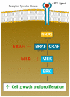
Oncogenic function of the MAPK signaling pathway. Genetic aberrations in this pathway are found in a vast majority of melanomas, with driver mutations in BRAF (V600E or V600K) occurring most often. The activation of BRAF results into phosphorylation of MEK and the activation of downstream MAP kinases like ERK. The pathway co-regulates tumor cell proliferation and cell survival; inhibition of BRAF downregulates the oncogenic function of MAPK signaling [33]. However, resistance to BRAF therapy is observed in about 50% of BRAF-mutated patients within 6–7 months after start of therapy. The CRAF-mediated reactivation of MAPK signaling pathway can be effectively blocked by the additional use of a MEK inhibitor (combined BRAF-MEK blockade), prolonging time to resistance development considerably. Consequently, anti-BRAF directed monotherapy is no longer used today. Figure adapted from Jenkins RW [67] and created byGellrich FF.
Figure 4
Figure 4
Proposed mechanisms of action of BEMPEG. After irreversible release of its six releasable polyethylene glycol (PEG) chains alternating its pharmacokinetic properties and its receptor binding, bempegaldesleukin is thought to expand and to activate CD8+ effector T cells and natural killer (NK) cells over T-regulatory cells (Tregs) [88]. The generation of active IL-2 conjugates with limited binding ability to the IL-2Rα subunit, thereby favoring the formation of dimeric βγ-IL-2 receptors (IL-2Rβγ; CD122), may explain its immunological activity investigated in vivo. Figure adapted from Charych D [94] and created by Gellrich FF, first published in J. Clin. Med. [23].

References

    1. Hanahan D., Weinberg R.A. The Hallmarks of Cancer. Cell. 2000;100:57–70. doi: 10.1016/S0092-8674(00)81683-9. - DOI - PubMed
    1. Hanahan D., Weinberg R.A. Hallmarks of Cancer: The next Generation. Cell. 2011;144:646–674. doi: 10.1016/j.cell.2011.02.013. - DOI - PubMed
    1. Miller A.J., Mihm M.C. Melanoma. N. Engl. J. Med. 2006;355:51–65. doi: 10.1056/NEJMra052166. - DOI - PubMed
    1. Davies M.A. The Role of the PI3K-AKT Pathway in Melanoma. Cancer J. 2012;18:142–147. doi: 10.1097/PPO.0b013e31824d448c. - DOI - PubMed
    1. Schadendorf D., Fisher D.E., Garbe C., Gershenwald J.E., Grob J.-J., Halpern A., Herlyn M., Marchetti M.A., McArthur G., Ribas A., et al. Melanoma. Nat. Rev. Dis Primers. 2015;1:15003. doi: 10.1038/nrdp.2015.3. - DOI - PubMed

LinkOut - more resources