Review
. 2021 Mar 24;22(7):3306.
doi: 10.3390/ijms22073306.

Islet Regeneration: Endogenous and Exogenous Approaches

Affiliations
Review

Islet Regeneration: Endogenous and Exogenous Approaches

Fiona M Docherty et al. Int J Mol Sci. .

Abstract

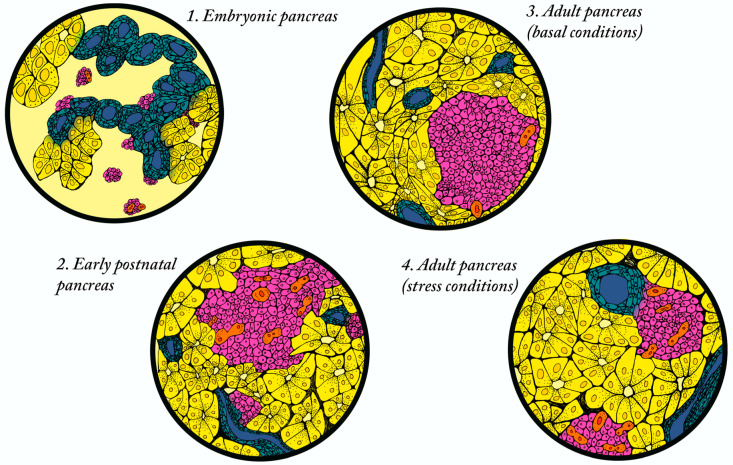
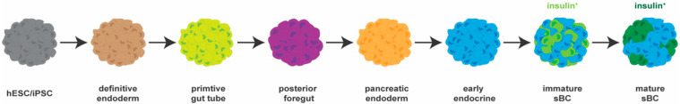
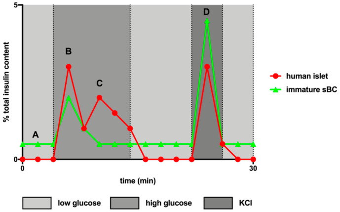
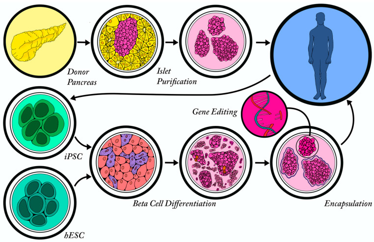
Both type 1 and type 2 diabetes are characterized by a progressive loss of beta cell mass that contributes to impaired glucose homeostasis. Although an optimal treatment option would be to simply replace the lost cells, it is now well established that unlike many other organs, the adult pancreas has limited regenerative potential. For this reason, significant research efforts are focusing on methods to induce beta cell proliferation (replication of existing beta cells), promote beta cell formation from alternative endogenous cell sources (neogenesis), and/or generate beta cells from pluripotent stem cells. In this article, we will review (i) endogenous mechanisms of beta cell regeneration during steady state, stress and disease; (ii) efforts to stimulate endogenous regeneration and transdifferentiation; and (iii) exogenous methods of beta cell generation and transplantation.

Keywords: beta cell; diabetes; islet; pancreas; regeneration.


Conflict of interest statement

The authors declare no conflict of interest. The funders had no role in the design of the study; in the collection, analyses, or interpretation of data; in the writing of the manuscript, or in the decision to publish the results.

Figures

Figure 1
Figure 1
Different mechanisms of endogenous beta cell regeneration exist throughout life. (1) Embryonic pancreas. During embryonic development, the beta cells primarily arise from the Neurogenin3 (NEUROG3)-expressing endocrine progenitor population located in epithelial ducts through a mechanism of neogenesis. (2) Early postnatal pancreas. In the first two weeks after birth, the beta cell population expands through a combination of neogenesis and replication of existing beta cells. (3) Adult pancreas in basal conditions. In the adult, a limited number of new beta cells form through replication. (4) Adult islet in stressed conditions. Adult beta cells exposed to physiological stresses (pregnancy, obesity, insulin resistance, damage) regenerate through both neogenesis (rare) and the replication of existing beta cells. Key: beta cells (pink); epithelial ducts (blue); replicating beta cells (orange); acinar cells (yellow).
Figure 2
Figure 2
Stem cell-derived beta-like cell differentiation protocols recapitulate developmental cues. Human pluripotent stem cells (hPSCs) are characterized by the expression of OCT4, NANOG and SOX2. The most successful beta cell differentiation programs recapitulate normal development. The first step includes a transition to a definitive endoderm (DE) cell fate that is driven by activation of the WNT and TGFβ pathways. After two days of exposure to the small molecules CHIR (WNT agonist) and Activin-A (TGFβ agonist), the cells express transcription factors specific to the definitive endoderm (DE) stage of development including SOX17. Next, the DE cells are differentiated towards the primitive gut tube (PGT) by the activation of the fibroblast growth factor (FGF) signaling pathway with KGF, a process that takes approximately three days. For the cells to activate the key pancreatic transcription factor, PDX1, and become specified to the posterior foregut (PF) they must receive cues that activate retinoic acid and hedgehog signaling and cues that inhibit Bone Morphogenetic Protein (BMP) signaling. At this stage, the cells are primed to be differentiated into any cell type in the pancreas and will become more fate restricted with time. The PDX1 positive PF cells are then differentiated into pancreatic endoderm (PE) cells that express PDX1 and NKX6.1 by activation of the FGF signaling pathway with EGF and KGF. These cells then undergo endocrine differentiation, during which they are exposed to signals that direct them to pancreatic beta cell fate while blocking differentiation to other pancreatic cell types. The key factors involved at this stage block TGFβ, BMP and Notch signaling. As they progress from the PE stage, the cells transiently express transcription factor NGN3 and then become NKX2.2 expressing early endocrine cells (EEs). NKX2.2 induction has been shown in 90–95% of cells at this stage demonstrating that early endocrine differentiation can be performed at very high efficiency [151]. After ~ ten days of endocrine differentiation, the cells express the key beta cell markers and begin to produce the insulin hormone, however, they are not yet considered functional as they do not regulate insulin secretion in response to glucose stimulation. A further five–seven days of maturation in a minimal media is required to allow the cells to become functional immature stem cell-derived beta-like cells (sBCs). Additional maturation steps, such as in vitro aggregation or transplantation into mice or humans, is required to achieve functionally mature sBCs. iPSC: induced pluripotent stem cell; hESC: human embryonic stem cells.
Figure 3
Figure 3
Dynamic glucose stimulated insulin secretion profiles depend on the maturation state of beta cells. Schematic representation of idealized dynamic glucose stimulated insulin secretion (dGSIS) profiles for cadaveric human islets 24 h post retrieval and immature sBCs obtained by perifusion. The values are presented as a percentage of total insulin content of the cluster of cells recovered following perifusion. Low glucose is 0.5 mM glucose in Krebs-Ringer Bicarbonate (KRB) buffer, high glucose is 16.7 mM glucose in KRB buffer and KCl is 30 mM KCl + 16.7 mM glucose in KRB buffer. A, Immature sBCs present with an increased glucose secretion in basal glucose. B, Immature sBCs respond to high glucose though often not to the same degree as human islets. C, Immature sBCs lack the characteristic second phase response to high glucose observed in human islets. D, Immature sBCs often show an exaggerated or so-called “uncontrolled” response to membrane depolarization with KCl; while human islets respond by secreting insulin at a level similar to their maximum response to high glucose.
Figure 4
Figure 4
Exogenous sources of beta cells. Exogenous beta cells can be purified from cadaveric beta cells isolated from a donor pancreas and transplanted into a T1D patient following the Edmonton protocol. Alternatively, terminally differentiated cells can be isolated from an individual and reprogrammed into iPSCs, followed by directed differentiation in vitro to form stem cell-derived beta-like cells (sBCs). In addition, hESCs can be differentiated to form non-patient specific sBCs. The sBCs from both iPSCs and hESCs can be genetically manipulated or encapsulated to help them evade the patient’s immune system following transplantation.

References

    1. Deng S., Vatamaniuk M., Huang X., Doliba N., Lian M.M., Frank A., Velidedeoglu E., Desai N.M., Koeberlein B., Wolf B., et al. Structural and functional abnormalities in the islets isolated from type 2 diabetic subjects. Diabetes. 2004;53:624–632. doi: 10.2337/diabetes.53.3.624. - DOI - PubMed
    1. Marselli L., Piron A., Suleiman M., Colli M.L., Yi X., Khamis A., Carrat G.R., Rutter G.A., Bugliani M., Giusti L., et al. Persistent or Transient Human beta Cell Dysfunction Induced by Metabolic Stress: Specific Signatures and Shared Gene Expression with Type 2 Diabetes. Cell Rep. 2020;33:108466. doi: 10.1016/j.celrep.2020.108466. - DOI - PubMed
    1. Ritzel R.A., Butler A.E., Rizza R.A., Veldhuis J.D., Butler P.C. Relationship between beta-cell mass and fasting blood glucose concentration in humans. Diabetes Care. 2006;29:717–718. doi: 10.2337/diacare.29.03.06.dc05-1538. - DOI - PubMed
    1. Linnemann A.K., Baan M., Davis D.B. Pancreatic beta-cell proliferation in obesity. Adv. Nutr. 2014;5:278–288. doi: 10.3945/an.113.005488. - DOI - PMC - PubMed
    1. Wright J.J., Saunders D.C., Dai C., Poffenberger G., Cairns B., Serreze D.V., Harlan D.M., Bottino R., Brissova M., Powers A.C. Decreased pancreatic acinar cell number in type 1 diabetes. Diabetologia. 2020;63:1418–1423. doi: 10.1007/s00125-020-05155-y. - DOI - PMC - PubMed

LinkOut - more resources