Case Reports
. 2021 Mar 24;57(4):302.
doi: 10.3390/medicina57040302.

COVID-19 and Pneumocystis jirovecii Pulmonary Coinfection-The First Case Confirmed through Autopsy

Affiliations
Case Reports

COVID-19 and Pneumocystis jirovecii Pulmonary Coinfection-The First Case Confirmed through Autopsy

Ionuț Isaia Jeican et al. Medicina (Kaunas). .

Abstract

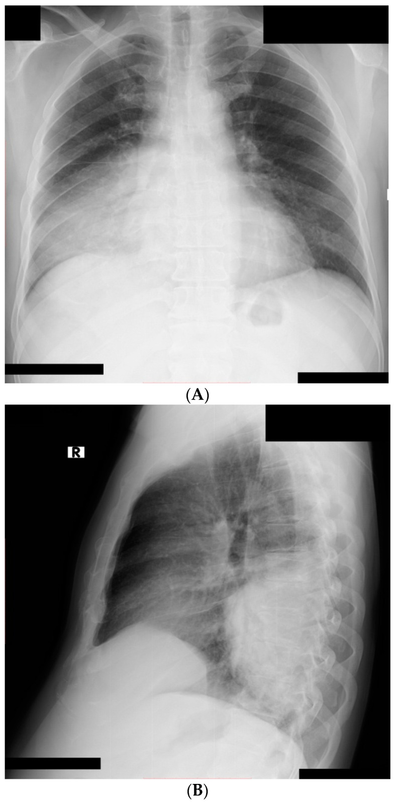
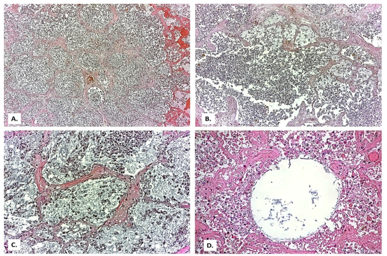
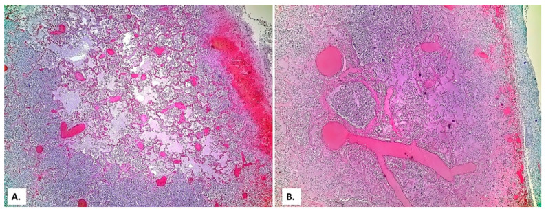
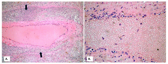
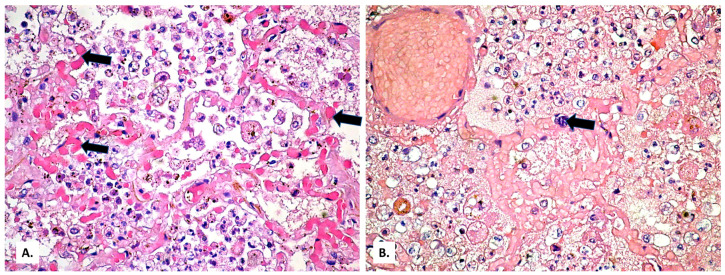
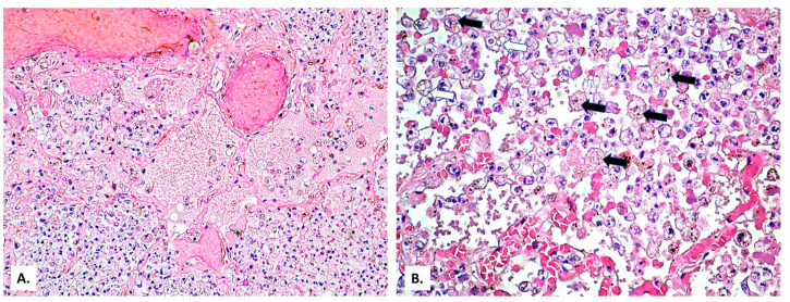
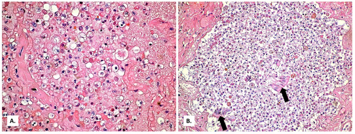
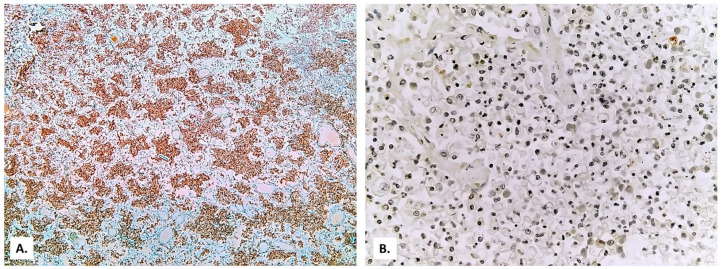
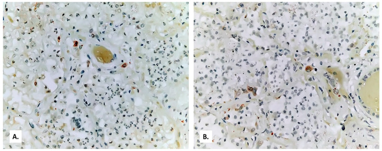
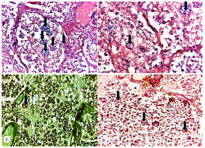
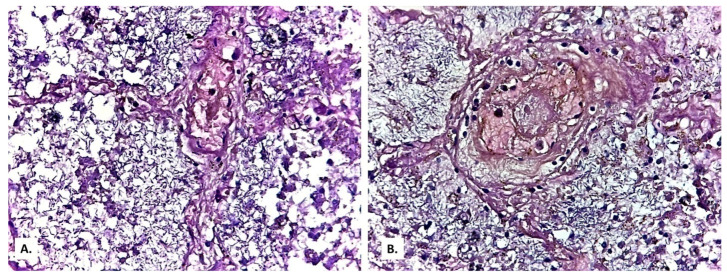
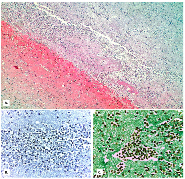
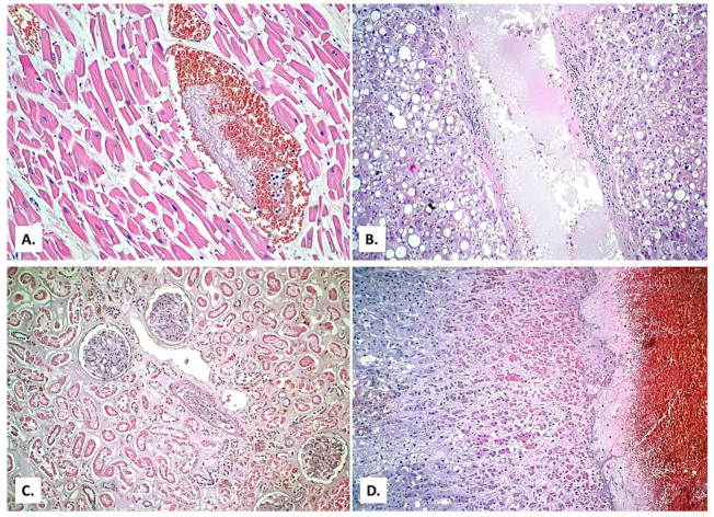
Background: Establishing the diagnosis of COVID-19 and Pneumocystisjirovecii pulmonary coinfection is difficult due to clinical and radiological similarities that exist between the two disorders. For the moment, fungal coinfections are underestimated in COVID-19 patients. Case presentation: We report the case of a 52-year-old male patient, who presented to the emergency department for severe dyspnea and died 17 h later. The RT-PCR test performed at his admission was negative for SARS-CoV-2. Retesting of lung fragments collected during autopsy revealed a positive result for SARS-CoV-2. Histopathological examination showed preexisting lesions, due to comorbidities, as well as recent lesions: massive lung thromboses, alveolar exudate rich in foam cells, suprapleural and intra-alveolar Pneumocystisjirovecii cystic forms, and bilateral adrenal hemorrhage. Conclusion: COVID-19 and P.jirovecii coinfection should be considered, particularly in critically ill patients, and we recommend the systematic search for P. jirovecii in respiratory samples.

Keywords: COVID-19; Pneumocystis jirovecii; SARS-CoV-2; autopsy; coinfection; pneumonia.


Conflict of interest statement

The authors declare no conflict of interest.

Figures

Figure 1
Figure 1
Chest X-ray: right pulmonary condensation syndrome ((A) postero-anterior view; (B) latero-lateral view).
Figure 2
Figure 2
(A) General appearance of diffusely thickened alveolar septa (HE × 40). (B) Emphysema bubble surrounded by thick fibrous septa (HE × 100). (C,D) Thickened septa with myofibroblasts (HE × 200). In all images, the presence of abundant intra alveolar inflammatory exudate can be observed.
Figure 3
Figure 3
(A) Extensive vascular thromboses in the venous territory and a pulmonary edema focus below a subpleural hemorrhage area. (B) Peribronchial pulmonary artery thromboses (a bronchial lumen is seen in the center of the image). Severe stasis with subpleural hemorrhage and the presence of basophilic exudate on the surface of the pleura (HE × 40).
Figure 4
Figure 4
(A) Thrombosed arterial vessel, dilated perivascular lymphatics (arrows) filled with a cell-rich lymph fluid (HE × 40). (B) The same vessel, at a high magnification, allows evidencing rare lymphocytes among the endothelial cells (HE × 400).
Figure 5
Figure 5
(A) Hyaline microthrombi (formed by fibrin) in the alveolar capillaries (arrows). (B) Basophilic microthrombus (formed by platelets) in the alveolar capillaries (arrows) (HE × 200).
Figure 6
Figure 6
(A) Zonally different aspects of alveolar exudate (HE × 100). (B) Alveolar exudate rich in foamy macrophages (black arrows) and cystic forms (white arrows) (HE × 400).
Figure 7
Figure 7
(A) Alveolus presenting a thickened wall with fibroblasts and abundant foamy macrophage content, on a background of granular eosinophilic material that fills the alveolus (HE × 400). (B) The exudate abundant in foamy macrophages also reaches the bronchial lumen which shows desquamated epithelium (arrows) (HE × 100).
Figure 8
Figure 8
(A) Immunolabeling with leukocyte common antigen (CD45) (×40). (B) Immunolabeling with CD3 (×200).
Figure 9
Figure 9
Immunolabeling for iNOS positive only in some macrophages ((A) microscopic field 1; (B) microscopic field 2).
Figure 10
Figure 10
(A,B) Cystic forms positive by Giemsa stain, (C) Grocott-Gomori’s methenamine silver stain, and (D) PAS stain (black arrows—full cystic forms; white arrow—empty cystic form).
Figure 11
Figure 11
Filamentous bacterial invasion (Giemsa × 400) ((A) microscopic field 1; (B) microscopic field 2).
Figure 12
Figure 12
(A) Eosinophilic filaments of fibrin at the contact with the pleura, covered by a cell-rich basophilic material (HE × 100). (B) Giemsa stain and (C) Grocott-Gomori’s methenamine silver stain are positive.
Figure 13
Figure 13
(A) Hypertrophic and fragmented myocardial fibers with a microthrombus in a venous vessel (HE × 400). (B) Liver with severe alcoholic steatosis and inflammation of the suprahepatic vein wall (HE × 100). (C) Kidney with tubular necrosis (HE × 200). (D) Adrenal medullary hemorrhage (HE × 40).

Similar articles

Cited by

References

    1. Jin M., Shi N., Wang M., Shi C., Lu S., Chang Q., Sha S., Lin Y., Chen Y., Zhou H., et al. CD45: A critical regulator in immune cells to predict severe and non-severe COVID-19 patients. Aging. 2020;12:19867–19879. doi: 10.18632/aging.103941. - DOI - PMC - PubMed
    1. Cao X. COVID-19: Immunopathology and its implications for therapy. Nat. Rev. Immunol. 2020;20:269–270. doi: 10.1038/s41577-020-0308-3. - DOI - PMC - PubMed
    1. Wiersinga W.J., Rhodes A., Cheng A.C., Peacock S.J., Prescott H.C. Pathophysiology, Transmission, Diagnosis, and Treatment of Coronavirus Disease 2019 (COVID-19): A Review. JAMA. 2020;324:782–793. doi: 10.1001/jama.2020.12839. - DOI - PubMed
    1. Bhat P., Noval M., Doub J.B., Heil E. Concurrent COVID-19 and Pneumocystis jirovecii pneumonia in a severely immunocompromised 25-year-old patient. Int. J. Infect. Dis. 2020;99:119–121. doi: 10.1016/j.ijid.2020.07.061. - DOI - PMC - PubMed
    1. Messina F.A., Marin E., Caceres D.H., Romero M., Depardo R., Priarone M.M., Rey L., Vázquez M., Verweij P.E., Chiller T.M., et al. Coronavirus Disease 2019 (COVID-19) in a Patient with Disseminated Histoplasmosis and HIV—A Case Report from Argentina and Literature Review. J. Fungi. 2020;6:275. doi: 10.3390/jof6040275. - DOI - PMC - PubMed

Publication types

MeSH terms

LinkOut - more resources