. 2021 Mar 24;10(4):722.
doi: 10.3390/cells10040722.

CXCL12-Abundant Reticular (CAR) Cells Direct Megakaryocyte Protrusions across the Bone Marrow Sinusoid Wall

Affiliations

CXCL12-Abundant Reticular (CAR) Cells Direct Megakaryocyte Protrusions across the Bone Marrow Sinusoid Wall

Nicole Wagner et al. Cells. .

Abstract

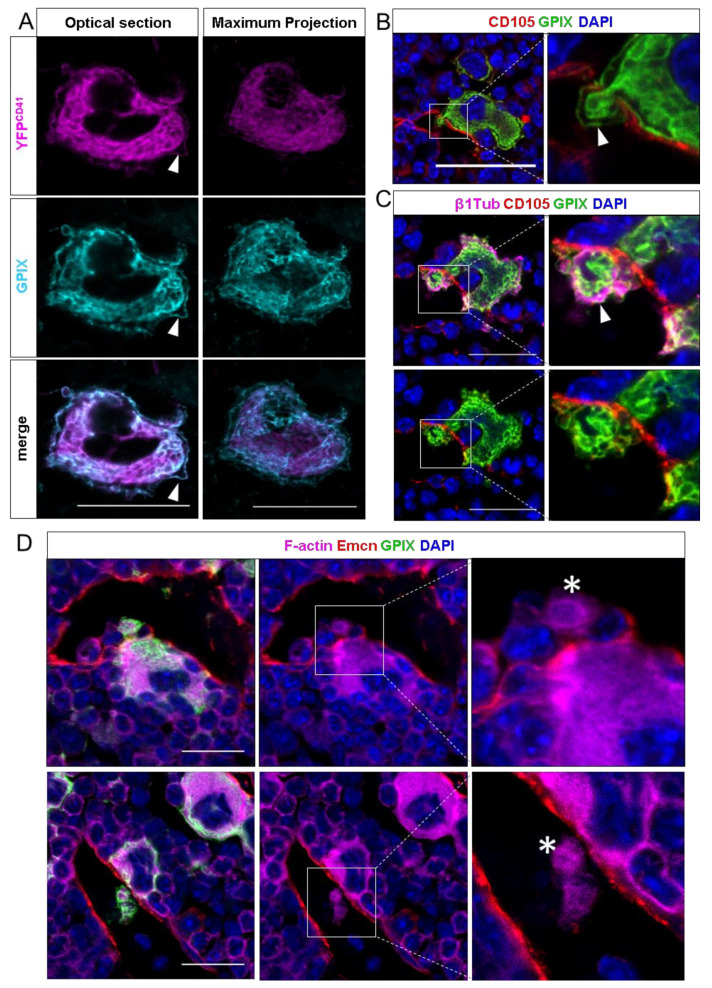
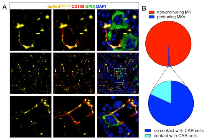
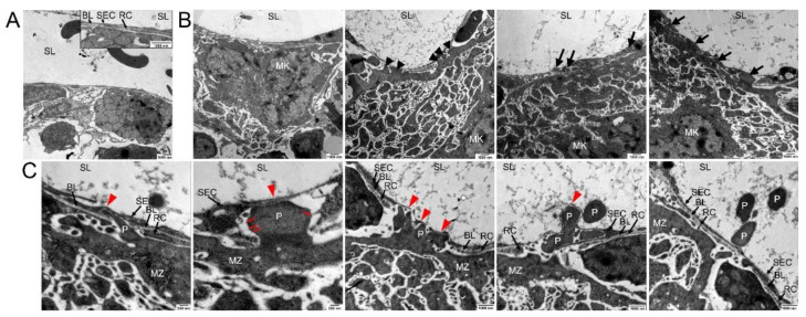
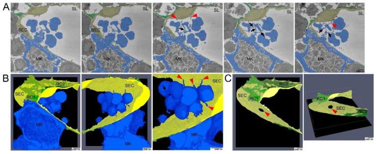
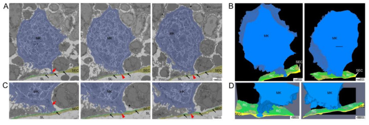
Megakaryocytes (MKs) release platelets into the lumen of bone marrow (BM) sinusoids while remaining to reside within the BM. The morphogenetic events of this complex process are still not fully understood. We combined confocal laser scanning microscopy with transmission and serial block-face scanning electron microscopy followed by 3D-reconstruction on mouse BM tissue sections. These analyses revealed that MKs in close vicinity to BM sinusoid (BMS) wall first induce the lateral retraction of CXCL12-abundant reticular (CAR) cells (CAR), followed by basal lamina (BL) degradation enabling direct MK-sinusoidal endothelial cells (SECs) interaction. Subsequently, an endothelial engulfment starts that contains a large MK protrusion. Then, MK protrusions penetrate the SEC, transmigrate into the BMS lumen and form proplatelets that are in direct contact to the SEC surface. Furthermore, such processes are induced on several sites, as observed by 3D reconstructions. Our data demonstrate that MKs in interaction with CAR-cells actively induce BMS wall alterations, including CAR-cell retraction, BL degradation, and SEC engulfment containing a large MK protrusion. This results in SEC penetration enabling the migration of MK protrusion into the BMS lumen where proplatelets that are adherent to the luminal SEC surface are formed and contribute to platelet release into the blood circulation.

Keywords: CXCL12-abundant reticular (CAR)-cells; megakaryocytes; microvasculature.


Conflict of interest statement

The authors declare no conflict of interest.

Figures

Figure 1
Figure 1
Confocal images of murine femur sections. (A) CD41wt/eYFP MKs were co-stained with GPIX. Optical sections show a strong signal overlap for the DMS, but less for the plasma membrane. In maximum projections, the plasma membrane staining becomes less visible. (B) Detection of intravasating megakaryocytes by immunohistochemical staining against GPIX and the endothelial marker endoglin (CD105). MKs enter the sinusoidal lumen as large protrusions and maintain a marginal zone (MZ) while crossing the endothelial lining. (C) Co-staining of vessels with GPIX and β1-tubulin. The MK-specific microtubule isoforms fill the MZ, while protruding the endothelial lining of the sinusoid. (D) Visualization of the F-actin cytoskeleton with sinusoids, stained by an additional vessel-marker endomucin (Emcn) and MKs. Large MK protrusions in the vessel lumen are highly actin-enriched, displaying the involvement of the actin-cytoskeleton in the transendothelial passage of MK protrusions. White arrows indicate the MZ, actin-rich proplatelets are marked with asteriscs. Scale bars 20 μm.
Figure 2
Figure 2
(A) dsRed staining of mouse femur sections shows a reticulated pattern (middle row). Resting MKs are in contact with CAR cells (bottom row); the endothelial lining shows pores only when MKs protrude. In these cases, CAR cells were absent or had retracted. (B) Quantification of MK-CAR cell contact. About 2% of MKs are protruding the vessel wall; only 25% of them are somewhat in contact with the dsRed expressing CAR cell. Scale bars 20 μm.
Figure 3
Figure 3
Transmission electron microscopical analysis of megakaryocyte (MK) transmural passage. (A) TEM image of components of the blood-BM-barrier, consisting of sinusoidal endothelial cells (SE) which are partially underlined by a basal lamina (BL) and covered by reticular cells (RC) to a large extent. No detection of any preformed pores in the sinusoidal endothelial cells was observed. Sinusoidal lumen (SL). (B) TEM images of approaching MKs. Before platelet protrusion, MKs approach the continuous endothelium with their MZ at various points (arrowheads) followed by invagination of MK protrusions into the continuous sinus endothelium (arrows). (C) TEM images of different steps of platelet (P) protrusion at higher magnification. At MK/SE contact sites (red arrowhead), MK protrusions provoke local retraction of reticular cells and disintegration of the basal lamina, followed by invagination of MK protrusions into the continuous sinus endothelium, massive stretching of SEs and subsequently rupture of the sinus endothelium and transcellular pore formation. Fine filaments are visible that anchor the protrusion to the SEC (red arrows).
Figure 4
Figure 4
Serial block face scanning electron microscopical analysis of MK transmural passage (A) Selected images from single SBF-SEM slices showing a megakaryocyte (MK) (blue) adjacent to the sinusoidal endothelial cells (SECs) (yellow) and (pro)platelets that are still interconnected via small cellular bridges (black arrows) and in contact with SECs (red arrowhead). MK protrusions provoke local retraction of reticular cells (RCs) (green). (B) Large-volume reconstruction of the SBF-SEM slices presented in (A) showing the relationship between a MK protrusion (blue) and the SECs (yellow). Before release of mature platelets, (pro)platelets are still interconnected via small cellular bridges (black arrow) and they stay still in contact with SECs (red arrowheads). MK protrusions provoke local retraction of RCs (green). (C) Large-volume reconstruction of the SBF-SEM slices presented in (A) clearly demonstrates MK extensions to cross the blood-BM-barrier by trans-endothelial passage. Model of sinusoidal endothelial cells (yellow) and RCs (green) depicting the site of trans-endothelial passage (hole, read arrowhead) of a MK protrusion (blue) and the local retraction of RCs (green). SBF-SEM dataset: Reconstruction of a 5 µm volume: 168 sections, 10 nm × 10 nm × 30 nm voxel size. Bone marrow of the diaphysis region obtained from murine ulnae. Source data are available for this figure.
Figure 5
Figure 5
Electron microscopical analysis of local retraction of CAR-cells (reticular cells, RCs) and disintegration of the BL at distinct contact sites of megakaryocyte (MK) protrusions. (A) Selected images from single SBF-SEM slices showing a MK (blue) adjacent to the sinusoidal endothelial cells (SECs, yellow). MK protrusions provoke local retraction of RCs (green) at two distinct contact sites where they are associated with the endothelial cell layer. (B) Large-volume reconstruction of the SBF-SEM slices presented in (A) demonstrating the relationship between a MK protrusion (blue), RCs (black arrows) and SECs (yellow). MK protrusions provoke local retraction of RCs (green) at two distinct sides of SECs contact while these contact sides are still surrounded by RC processes. (C) Selected images from single SBF-SEM slices of (A) at higher magnification showing a MK protrusion in close association with the blood-BM-barrier. Although the MK protrusion is in close association with this barrier, RCs still cover the endothelial lining to a large extend (red arrow). RCs are retracted locally at two distinct sites where the MK protrusion is associated with the endothelial cell layer (red arrowheads) while the contact sides are still surrounded by reticular cell processes (black arrows). (D) Large-volume reconstruction of the SBF-SEM slices presented in (B) at higher magnification. SBF-SEM dataset: Reconstruction of a 3.9 μm volume: 132 sections, 10 nm × 10 nm × 30 nm voxel size. BM of the diaphysis region obtained from murine ulnae. Source data are available for this figure. Sinusoidal lumen (SL); Protrusion (P); Marginal zone (MZ).

Similar articles

Cited by

References

    1. Italiano J.E., Jr., Lecine P., Shivdasani R.A., Hartwig J.H. Blood platelets are assembled principally at the ends of proplatelet processes produced by differentiated megakaryocytes. J. Cell Biol. 1999;147:1299–1312. doi: 10.1083/jcb.147.6.1299. - DOI - PMC - PubMed
    1. De Bruyn P.P.H., Michelson S., Thomas T.B. Migration of Blood Cells of Bone Marrow through Sinusoidal Wall. J. Morphol. 1971;133:417. doi: 10.1002/jmor.1051330406. - DOI - PubMed
    1. Slayton W.B., Li X.M., Butler J., Guthrie S.M., Jorgensen M.L., Wingard J.R., Scott E.W. The role of the donor in the repair of the marrow vascular niche following hematopoietic stem cell transplant. Stem Cells. 2007;25:2945–2955. doi: 10.1634/stemcells.2007-0158. - DOI - PubMed
    1. Junt T., Schulze H., Chen Z., Massberg S., Goerge T., Krueger A., Wagner D.D., Graf T., Italiano J.E., Jr., Shivdasani R.A., et al. Dynamic visualization of thrombopoiesis within bone marrow. Science. 2007;317:1767–1770. doi: 10.1126/science.1146304. - DOI - PubMed
    1. Nishimura S., Nagasaki M., Kunishima S., Sawaguchi A., Sakata A., Sakaguchi H., Ohmori T., Manabe I., Italiano J.E., Ryu T., et al. IL-1 alpha induces thrombopoiesis through megakaryocyte rupture in response to acute platelet needs. J. Cell Biol. 2015;209:453–466. doi: 10.1083/jcb.201410052. - DOI - PMC - PubMed

Publication types

MeSH terms

Substances

LinkOut - more resources