. 2021 Mar 24;12(4):347.
doi: 10.3390/mi12040347.

A Portable Microfluidic System for Point-of-Care Detection of Multiple Protein Biomarkers

Affiliations

A Portable Microfluidic System for Point-of-Care Detection of Multiple Protein Biomarkers

Nan Li et al. Micromachines (Basel). .

Abstract

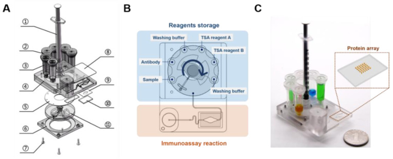
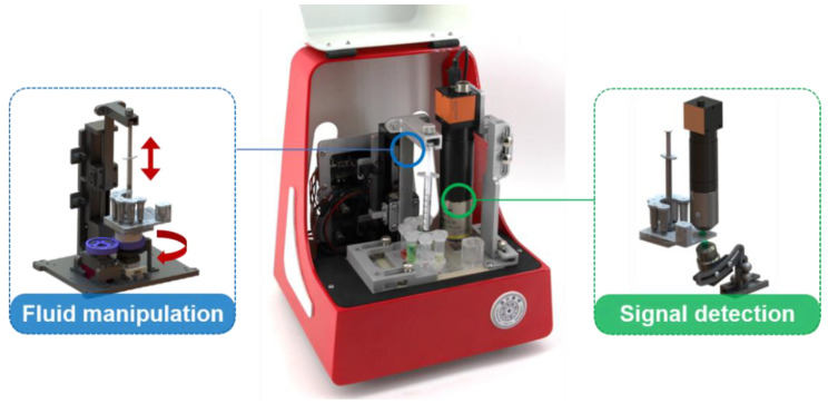
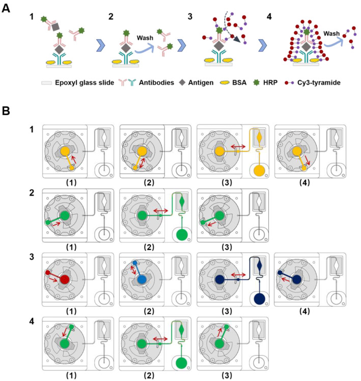
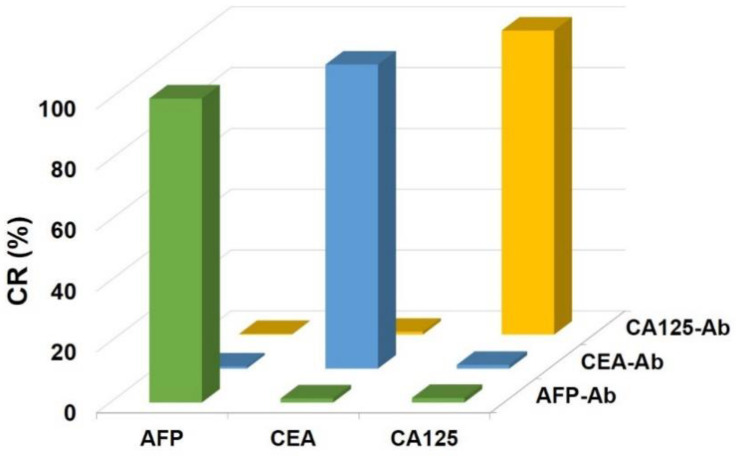
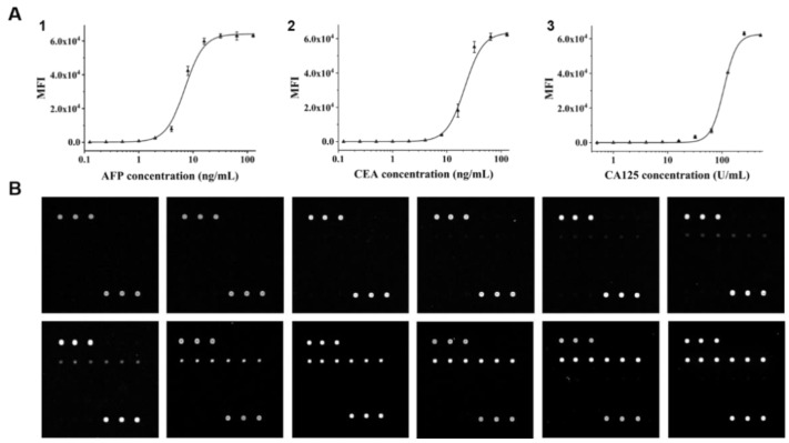
Protein biomarkers are indicators of many diseases and are commonly used for disease diagnosis and prognosis prediction in the clinic. The urgent need for point-of-care (POC) detection of protein biomarkers has promoted the development of automated and fully sealed immunoassay platforms. In this study, a portable microfluidic system was established for the POC detection of multiple protein biomarkers by combining a protein microarray for a multiplex immunoassay and a microfluidic cassette for reagent storage and liquid manipulation. The entire procedure for the immunoassay was automatically conducted, which included the antibody-antigen reaction, washing and detection. Alpha-fetoprotein (AFP), carcinoembryonic antigen (CEA) and carcinoma antigen 125 (CA125) were simultaneously detected in this system within 40 min with limits of detection of 0.303 ng/mL, 1.870 ng/mL, and 18.617 U/mL, respectively. Five clinical samples were collected and tested, and the results show good correlations compared to those measured by the commercial instrument in the hospital. The immunoassay cassette system can function as a versatile platform for the rapid and sensitive multiplexed detection of biomarkers; therefore, it has great potential for POC diagnostics.

Keywords: immunoassay; microarray; microfluidic cassette; multiplex measurement; point-of-care testing; protein biomarker.


Conflict of interest statement

The authors declare no conflict of interest.

Figures

Figure 1
Figure 1
Schematic of the cassette used for the immunoassay. (A) The three-dimensional illustration of the cassette in an exploded view, 1. Luer syringe; 2. lid; 3. reagents and waste storage chambers; 4. main body of the cassette; 5. polyurethane washer; 6. cassette base; 7. screws; 8. pressure-sensitive adhesive (PSA) cover slip; 9. double adhesive tape; 10. glass; 11. rotary valve. (B) Schematic layout of the microfluidic cassette indicating various features. The blue area represents the reagent storage module, and the yellow area represents the immunoassay reaction module. (C) Photo of the cassette. The yellow box shows the glass with a protein microarray.
Figure 2
Figure 2
The completed prototype of the supporting device with fluid manipulation and signal detection modules.
Figure 3
Figure 3
The cassette processing protocol. (A) Schematic showing the process of the immunoassay. (1) Antibody–antigen reaction; (2) washing; (3) horseradish peroxidase (HRP)-catalyzed signal amplification; (4) washing. The Cy3-tyramide molecule can be catalyzed by HRP under a H2O2 environment, and the activated tyramide can covalently bind to certain amino acid residues of the proximate proteins. (B) Illustration of the entire flow control system of the cassette: (1) the 200 μL serum sample is mixed with lyophilized antibody and then placed into the reaction chamber; (2) after completing the antibody–antigen reaction, the glass is washed with 800 μL washing buffer; (3) 200 μL TSA reagent A and 200 μL TSA reagent B are mixed prior to transfer into the reaction chamber; (4) the glass is washed with 800 μL washing buffer.
Figure 4
Figure 4
Cross-reactivities of the three pairs of antigens and antibodies.
Figure 5
Figure 5
Establishment of the standard curves. (A) Calibration curves for the immunoassays of tumor markers. (1) Standard curve for alpha-fetoprotein (AFP); (2) standard curve for carcinoembryonic antigen (CEA); (3) standard curve for carcinoma antigen 125 (CA125). The error bars show the standard deviations of triplicate measurements. (B) Representative fluorescence images of microarrays during detection of AFP with simulated standard samples. The concentrations of AFP were 0, 0.125, 0.25, 0.5, 1, 2, 4, 8, 16, 32, 64 and 128 ng/mL (from left to right and top to bottom).

References

    1. Allinson J.L. Clinical biomarker validation. Bioanalysis. 2018;10:957–968. doi: 10.4155/bio-2018-0061. - DOI - PubMed
    1. Biomarkers Definitions Working Group. Atkinson A.J., Jr., Colburn W.A., DeGruttola V.G., DeMets D.L., Downing G.J., Hoth D.F., Oates J.A., Peck C.C., Schooley R.T., et al. Biomarkers and surrogate endpoints: Preferred definitions and conceptual framework. Clin. Pharmacol. Ther. 2001;69:89–95. - PubMed
    1. Califf R.M. Biomarker definitions and their applications. Exp. Biol. Med. 2018;243:213–221. doi: 10.1177/1535370217750088. - DOI - PMC - PubMed
    1. Wulfkuhle J.D., Liotta L.A., Petricoin E.F. Proteomic applications for the early detection of cancer. Nat. Rev. Cancer. 2003;3:267–275. doi: 10.1038/nrc1043. - DOI - PubMed
    1. Kingsmore S.F. Multiplexed protein measurement: Technologies and applications of protein and antibody arrays. Nat. Rev. Drug Discov. 2006;5:310–320. doi: 10.1038/nrd2006. - DOI - PMC - PubMed