The Graphical Representation of Cell Count Representation: A New Procedure for the Diagnosis of Periprosthetic Joint Infections
- PMID: 33804988
- PMCID: PMC8063952
- DOI: 10.3390/antibiotics10040346
The Graphical Representation of Cell Count Representation: A New Procedure for the Diagnosis of Periprosthetic Joint Infections
Abstract
Aim: This study was designed to answer the question whether a graphical representation increase the diagnostic value of automated leucocyte counting of the synovial fluid in the diagnosis of periprosthetic joint infections (PJI).
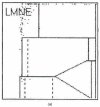
Material and methods: Synovial aspirates from 322 patients (162 women, 160 men) with revisions of 192 total knee and 130 hip arthroplasties were analysed with microbiological cultivation, determination of cell counts and assay of the biomarker alpha-defensin (170 cases). In addition, microbiological and histological analysis of the periprosthetic tissue obtained during the revision surgery was carried out using the ICM classification and the histological classification of Morawietz and Krenn. The synovial aspirates were additionally analysed to produce dot plot representations (LMNE matrices) of the cells and particles in the aspirates using the hematology analyser ABX Pentra XL 80.
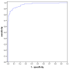
Results: 112 patients (34.8%) had an infection according to the ICM criteria. When analysing the graphical LMNE matrices from synovia cell counting, four types could be differentiated: the type "wear particles" (I) in 28.3%, the type "infection" (II) in 24.8%, the "combined" type (III) in 15.5% and "indeterminate" type (IV) in 31.4%. There was a significant correlation between the graphical LMNE-types and the histological types of Morawietz and Krenn (p < 0.001 and Cramer test V value of 0.529). The addition of the LMNE-Matrix assessment increased the diagnostic value of the cell count and the cut-off value of the WBC count could be set lower by adding the LMNE-Matrix to the diagnostic procedure.
Conclusion: The graphical representation of the cell count analysis of synovial aspirates is a new and helpful method for differentiating between real periprosthetic infections with an increased leukocyte count and false positive data resulting from wear particles. This new approach helps to increase the diagnostic value of cell count analysis in the diagnosis of PJI.
Keywords: cell count; diagnosis; leukocyte; periprosthetic joint infection.
Conflict of interest statement
The authors declare no conflict of interest.
Figures


Similar articles
-
Graphic type differentiation of cell count data for diagnosis of early and late periprosthetic joint infection: A new method.Technol Health Care. 2024;32(5):3669-3680. doi: 10.3233/THC-231006. Technol Health Care. 2024. PMID: 37980584 Free PMC article.
-
A New Graphic Type Differentiation of Cell Account Determination for Distinguishing Acute Periprosthetic Joint Infection from Hemarthrosis.Antibiotics (Basel). 2022 Sep 21;11(10):1284. doi: 10.3390/antibiotics11101284. Antibiotics (Basel). 2022. PMID: 36289943 Free PMC article.
-
False-Positive Automated Synovial Fluid White Blood Cell Counting Is a Concern for Both Hip and Knee Arthroplasty Aspirates.J Arthroplasty. 2020 Jun;35(6S):S304-S307. doi: 10.1016/j.arth.2020.01.060. Epub 2020 Jan 28. J Arthroplasty. 2020. PMID: 32113809
-
Diagnostic Accuracy of Serum, Synovial, and Tissue Testing for Chronic Periprosthetic Joint Infection After Hip and Knee Replacements: A Systematic Review.J Bone Joint Surg Am. 2019 Apr 3;101(7):635-649. doi: 10.2106/JBJS.18.00632. J Bone Joint Surg Am. 2019. PMID: 30946198
-
The role of synovial fluid analysis in the detection of periprosthetic hip and knee infections: a systematic review and meta-analysis.Int Orthop. 2018 May;42(5):983-994. doi: 10.1007/s00264-018-3865-3. Epub 2018 Mar 9. Int Orthop. 2018. PMID: 29523955
Cited by
-
Antibiotics in Orthopedic Infections.Antibiotics (Basel). 2021 Oct 25;10(11):1297. doi: 10.3390/antibiotics10111297. Antibiotics (Basel). 2021. PMID: 34827235 Free PMC article.
-
Graphic type differentiation of cell count data for diagnosis of early and late periprosthetic joint infection: A new method.Technol Health Care. 2024;32(5):3669-3680. doi: 10.3233/THC-231006. Technol Health Care. 2024. PMID: 37980584 Free PMC article.
-
A New Graphic Type Differentiation of Cell Account Determination for Distinguishing Acute Periprosthetic Joint Infection from Hemarthrosis.Antibiotics (Basel). 2022 Sep 21;11(10):1284. doi: 10.3390/antibiotics11101284. Antibiotics (Basel). 2022. PMID: 36289943 Free PMC article.
-
Differential synovial fluid white blood cell count for the diagnosis of chronic peri-prosthetic joint infection - a systematic review and meta-analysis.J Bone Jt Infect. 2025 May 14;10(3):165-184. doi: 10.5194/jbji-10-165-2025. eCollection 2025. J Bone Jt Infect. 2025. PMID: 40385309 Free PMC article.
-
Diagnostics in Late Periprosthetic Infections-Challenges and Solutions.Antibiotics (Basel). 2024 Apr 11;13(4):351. doi: 10.3390/antibiotics13040351. Antibiotics (Basel). 2024. PMID: 38667027 Free PMC article. Review.
References
LinkOut - more resources
Full Text Sources
Other Literature Sources

