. 2021 Mar 24;11(4):267.
doi: 10.3390/life11040267.

The Telomeric Protein TRF2 Regulates Replication Origin Activity within Pericentromeric Heterochromatin

Affiliations

The Telomeric Protein TRF2 Regulates Replication Origin Activity within Pericentromeric Heterochromatin

Serge Bauwens et al. Life (Basel). .

Abstract

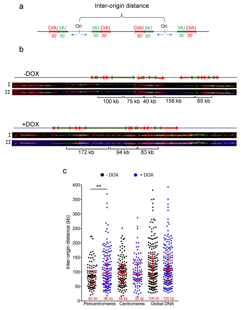
Heterochromatic regions render the replication process particularly difficult due to the high level of chromatin compaction and the presence of repeated DNA sequences. In humans, replication through pericentromeric heterochromatin requires the binding of a complex formed by the telomeric factor TRF2 and the helicase RTEL1 in order to relieve topological barriers blocking fork progression. Since TRF2 is known to bind the Origin Replication Complex (ORC), we hypothesized that this factor could also play a role at the replication origins (ORI) of these heterochromatin regions. By performing DNA combing analysis, we found that the ORI density is higher within pericentromeric satellite DNA repeats than within bulk genomic DNA and decreased upon TRF2 downregulation. Moreover, we showed that TRF2 and ORC2 interact in pericentromeric DNA, providing a mechanism by which TRF2 is involved in ORI activity. Altogether, our findings reveal an essential role for TRF2 in pericentromeric heterochromatin replication by regulating both replication initiation and elongation.

Keywords: ORC; TRF2; heterochromatin; pericentromeric DNA; replication; telomeres.


Conflict of interest statement

The authors declare no conflict of interest.

Figures

Figure 1
Figure 1
The ORC complex in pericentromeric regions. (a) Western blotting showing the expression of TRF2 in HeLa cells treated with doxycycline (DOX) for 5 days. The percentage of the remaining signal normalized to actin is shown at the bottom of the gel; (b) Slot blot showing the presence of ORC1 and ORC2 in pericentromeric regions. Membrane was hybridized with a radioactive satellite III DNA probe (left panel). Quantification of the signal obtained by normalizing the immunoprecipitated signal (IP) to that of the input is shown (right panel). Bars show SD of the mean of three biological replicates. (** p < 0.001; Mann–Whitney U test).
Figure 2
Figure 2
Origin of replication density in shTERF2-expressing HeLa cells. (a) Scheme showing the experimental setting used to measure the distance between two origins of replication (inter-origin distance; IOD); (b) Representative images of DNA combing at pericentromeres showing only the signal of IdU (green), IdU (red) top image (I) or with satellite III PNA probe signal (blue) bottom image (II). Green and red arrows indicate IdU and CldU labeling, respectively. IOD distance is shown below the images; (c) Quantification of IOD expressed in kilobases (kb) for HeLa cells expressing a doxycycline (DOX)-inducible shTERF2 system. Median length is shown at the bottom of the scatter plots. Bars represent the median +/− interquartile range (** p < 0.001; Mann–Whitney U test).
Figure 3
Figure 3
Effect of TRF2ΔB and TRF2ΔM on origin density. (a) Relevant TRF2 protein domains and functions; (b) TRF2 expression of HeLa cells treated for 5 days with doxycycline (DOX). One day after DOX was added to the media, the indicated lentivirus-expressing plasmids were added to the culture; (c) Quantification of inter-origin distances at pericentromeric regions expressed in kilobases (kb) for the conditions described in b. Median length is displayed at the bottom of the scatter plots. Bars represent the median +/− interquartile range (** p < 0.001; *** p < 0.0001; Mann–Whitney U test).

References

    1. Janssen A., Colmenares S.U., Karpen G.H. Heterochromatin: Guardian of the Genome. Annu. Rev. Cell Dev. Biol. 2018;34:265–288. doi: 10.1146/annurev-cellbio-100617-062653. - DOI - PubMed
    1. Mendez-Bermudez A., Giraud-Panis M.J., Ye J., Gilson E. Heterochromatin replication goes hand in hand with telomere protection. Nat. Struct. Mol. Biol. 2020;27:313–318. doi: 10.1038/s41594-020-0400-1. - DOI - PubMed
    1. Tazumi A., Fukuura M., Nakato R., Kishimoto A., Takenaka T., Ogawa S., Song J.H., Takahashi T.S., Nakagawa T., Shirahige K., et al. Telomere-binding protein Taz1 controls global replication timing through its localization near late replication origins in fission yeast. Genes Dev. 2012;26:2050–2062. doi: 10.1101/gad.194282.112. - DOI - PMC - PubMed
    1. Zofall M., Smith D.R., Mizuguchi T., Dhakshnamoorthy J., Grewal S.I.S. Taz1-Shelterin Promotes Facultative Heterochromatin Assembly at Chromosome-Internal Sites Containing Late Replication Origins. Mol. Cell. 2016;62:862–874. doi: 10.1016/j.molcel.2016.04.034. - DOI - PMC - PubMed
    1. Zimmermann M., Kibe T., Kabir S., de Lange T. TRF1 negotiates TTAGGG repeatassociated replication problems by recruiting the BLM helicase and the TPP1/POT1 repressor of ATR signaling. Genes Dev. 2014;28:2477–2491. doi: 10.1101/gad.251611.114. - DOI - PMC - PubMed

LinkOut - more resources