. 2021 Mar 24;13(4):542.
doi: 10.3390/v13040542.

The Change P82L in the Rift Valley Fever Virus NSs Protein Confers Attenuation in Mice

Affiliations

The Change P82L in the Rift Valley Fever Virus NSs Protein Confers Attenuation in Mice

Belén Borrego et al. Viruses. .

Abstract

Rift Valley fever virus (RVFV) is a mosquito-borne bunyavirus that causes an important disease in ruminants, with great economic losses. The infection can be also transmitted to humans; therefore, it is considered a major threat to both human and animal health. In a previous work, we described a novel RVFV variant selected in cell culture in the presence of the antiviral agent favipiravir that was highly attenuated in vivo. This variant displayed 24 amino acid substitutions in different viral proteins when compared to its parental viral strain, two of them located in the NSs protein that is known to be the major virulence factor of RVFV. By means of a reverse genetics system, in this work we have analyzed the effect that one of these substitutions, P82L, has in viral attenuation in vivo. Rescued viruses carrying this single amino acid change were clearly attenuated in BALB/c mice while their growth in an interferon (IFN)-competent cell line as well as the production of interferon beta (IFN-β) did not seem to be affected. However, the pattern of nuclear NSs accumulation was modified in cells infected with the mutant viruses. These results highlight the key role of the NSs protein in the modulation of viral infectivity.

Keywords: PXXP motifs; Rift Valley fever virus; interferon antagonist; non-structural NSs protein; nuclear filaments.


Conflict of interest statement

The authors declare no conflict of interest.

Figures
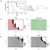
Figure 1
Figure 1
Analysis of the in vivo pathogenicity of the rZH548-P82L mutant viruses in BALB/c mice. 9–18-week-old male mice (n = 5–7, equally distributed) were inoculated IP with 500 plaque-forming units (pfus) of the indicated viruses and both rZH548-P82L clones, 2B7 and 3VB5. Wild-type rZH548 (red) and rZH548ΔNSs::GFP (labeled as, rZHΔNSs/GFP, green) viruses were included as controls for virulence and attenuation, respectively. Animals were monitored up to 18 days. (A) Survival rates and (BE) morbidity upon challenge with the indicated viruses. The graph represents the clinical status of each mouse: D (dead/euthanized): black bars; S (signs-sick), hatched bars; H (healthy), grey bars. The animal within the group rZH548ΔNSs::GFP euthanized at day 4 pi was excluded from the survival analysis.
Figure 2
Figure 2
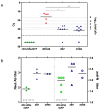
Viremia and seroconversion after inoculation with the rZH548-P82L mutant viruses. (A) Viremia. RT-qPCR on EDTA blood samples collected at day 3 pi. Samples giving a Cq (quantification cycle) value under the detection level of the assay (37) are arbitrarily represented as 45 and were excluded from the statistical analysis. The correlation of Cq data with pfu equivalents is indicated in the right Y axis. (B) Antibody responses in survivor mice at day 18 pi. Titers are expressed as the dilution of serum (log10) rendering a reduction in infectivity of 50% in a microneutralization assay (left Y-axis; closed symbols), and last dilution of serum (log10) giving an OD reading at 450 nm over 1.0 in anti-N ELISA (right Y-axis; open symbols). Each symbol corresponds to an individual mouse. For neutralization, only n = 3 samples were available for rZH548ΔNSs::GFP and 2B7. * p ≤ 0.05, ** p ≤ 0.001.
Figure 3
Figure 3
Localization and filament formation of wt and mutant NSs proteins. Vero cells were infected with rZH548 and the two rZH548-P82L mutants at a MOI of 1. At 6 (panel A) and 24 (panel B) hours pi, cells were fixed and subjected to indirect immunofluorescence with the anti-NSs monoclonal antibody 5C3A1B2. Nuclei were stained with DAPI. For each virus and time pi, 2 images with different magnification are shown as indicated. AS denotes Zeiss Airyscan 2D superresolution mode. Red arrows in panel A point to cells infected with the rZH548-P82L viruses where nuclear filaments could be detected. Scale bars: 20 µM (upper panel B), 10 µM (upper panel A and lower panel B); 5 µM (lower panel A).
Figure 4
Figure 4
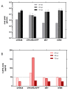
Growth of rZH548-P82L mutants on HEK293T cells and IFN-β production. HEK293T cells were infected at a MOI of 0.05 with the indicated viruses. At 24, 48 and 72 hpi, supernatants were collected and titrated on Vero cells (panel A) and analyzed for IFN-β production by ELISA (panel B). The limit of detection of this ELISA was established at 50 pg/mL (see Materials and Methods). The sample corresponding to rZH548 at 72 hpi was not analyzed in ELISA.
Figure 5
Figure 5
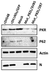
Degradation of protein kinase R (PKR) and p62 in rZH548-P82L infected cells. HEK293T cells were infected at a MOI of 1 with the indicated viruses. Cells were harvested at 20 hpi and analyzed by Western blot using anti-PKR (B-10), anti-p62 (H10) mouse monoclonal antibodies, anti RVFV-N mAb 2B1 and anti-actin antibody as primary antibodies. Samples loaded correspond to 106 cells.

References

    1. Ikegami T., Makino S. The pathogenesis of Rift Valley fever. Viruses. 2011;3:493–519. doi: 10.3390/v3050493. - DOI - PMC - PubMed
    1. Rolin A.I., Berrang-Ford L., Kulkarni M.A. The risk of Rift Valley fever virus introduction and establishment in the United States and European Union. Emerg. Microbes Infect. 2013;2:e81. doi: 10.1038/emi.2013.81. - DOI - PMC - PubMed
    1. Boshra H., Lorenzo G., Busquets N., Brun A. Rift valley fever: Recent insights into pathogenesis and prevention. J. Virol. 2011;85:6098–6105. doi: 10.1128/JVI.02641-10. - DOI - PMC - PubMed
    1. Kortekaas J. One Health approach to Rift Valley fever vaccine development. Antiviral Res. 2014;106:24–32. doi: 10.1016/j.antiviral.2014.03.008. - DOI - PubMed
    1. Faburay B., LaBeaud A.D., McVey D.S., Wilson W.C., Richt J.A. Current Status of Rift Valley Fever Vaccine Development. Vaccines. 2017;5:29. doi: 10.3390/vaccines5030029. - DOI - PMC - PubMed

Publication types

MeSH terms