Review
. 2021 Mar 24;22(7):3337.
doi: 10.3390/ijms22073337.

Pseudomonas Flagella: Generalities and Specificities

Affiliations
Review

Pseudomonas Flagella: Generalities and Specificities

Mathilde Bouteiller et al. Int J Mol Sci. .

Abstract

Flagella-driven motility is an important trait for bacterial colonization and virulence. Flagella rotate and propel bacteria in liquid or semi-liquid media to ensure such bacterial fitness. Bacterial flagella are composed of three parts: a membrane complex, a flexible-hook, and a flagellin filament. The most widely studied models in terms of the flagellar apparatus are E. coli and Salmonella. However, there are many differences between these enteric bacteria and the bacteria of the Pseudomonas genus. Enteric bacteria possess peritrichous flagella, in contrast to Pseudomonads, which possess polar flagella. In addition, flagellar gene expression in Pseudomonas is under a four-tiered regulatory circuit, whereas enteric bacteria express flagellar genes in a three-step manner. Here, we use knowledge of E. coli and Salmonella flagella to describe the general properties of flagella and then focus on the specificities of Pseudomonas flagella. After a description of flagellar structure, which is highly conserved among Gram-negative bacteria, we focus on the steps of flagellar assembly that differ between enteric and polar-flagellated bacteria. In addition, we summarize generalities concerning the fuel used for the production and rotation of the flagellar macromolecular complex. The last part summarizes known regulatory pathways and potential links with the type-six secretion system (T6SS).

Keywords: Pseudomonas; T6SS; flagella; flagellar crosstalk.


Conflict of interest statement

The authors declare no conflicts of interest. The funders had no role in the writing of the manuscript.

Figures
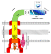
Figure 1
Figure 1
Schematic representation of the structure of the flagellar apparatus. IM, PG, and OM correspond to the inner membrane, peptidoglycan layer, and outer membrane, respectively. The flagellar membrane complex is composed of the basal body (in red), flagellar type 3 secretion system (in orange), and rod structure (in yellow). The flagellar filament (in various shades of blue) is connected to the hook (in green) by two junction proteins (in white). The flagellin cap (in purple) is the only cap protein that remains attached to the flagellar structure. The stator complex (in pink) is anchored to the peptidoglycan layer and interacts with the C-Ring to generate flagellar rotation.
Figure 2
Figure 2
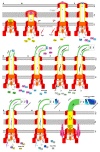
Schematic representation of flagellar assembly in nine arbitrary steps. IM: inner membrane, PG: peptidoglycan, OM: outer membrane. (A) Formation of the membrane complex and proximal rod. (B) Rod extension and P- and L-ring formation. (C) Hook cap protein insertion. (D) Hook extension. (E) Hook completion, substrate specificity switch, and FlgM secretion. (F) Junction protein addition. (G) Flagellin cap insertion. (H) Extension of the flagellin filament. (I) Insertion of the stator complex.
Figure 3
Figure 3
Schematic representation of the specifics of flagellar assembly in Salmonella/E. coli and Pseudomonas. Hierarchical transcription occurs in a three-step manner in Salmonella and E. coli (A) and in a four-step manner in Pseudomonas (B). Each box summarizes the production of a nascent flagellar structure and regulator proteins during expression of the corresponding class. Transcription is represented by the dotted arrows topped by the corresponding transcription factors.
Figure 4
Figure 4
Regulation of flagella in Pseudomonas. The assembly and function of flagella in Pseudomonas are regulated by various mechanisms, such as (A) chemotaxis, (B) temperature, (C) two-component systems, (D) cAMP concentration, (E) c-di-GMP concentration, and (F) T6SS. Quorum sensing (QS) (represented in a green bubble) also appears to be involved but no data on the regulation of Pseudomonas flagella by QS are available. The key elements and regulators of flagella are written in blue. Black arrows indicate positive regulation, dotted arrows indicate potential positive regulation, double arrows indicate an interaction, dotted double arrows correspond to a potential interaction, blunt red lines represent negative regulation, and question marks indicate an unknown mechanism or interaction.

References

    1. Soutourina O.A., Bertin P.N. Regulation Cascade of Flagellar Expression in Gram-Negative Bacteria. FEMS Microbiol. Rev. 2003;27:505–523. doi: 10.1016/S0168-6445(03)00064-0. - DOI - PubMed
    1. Wang F., Burrage A.M., Postel S., Clark R.E., Orlova A., Sundberg E.J., Kearns D.B., Egelman E.H. A Structural Model of Flagellar Filament Switching across Multiple Bacterial Species. Nat. Commun. 2017;8:960. doi: 10.1038/s41467-017-01075-5. - DOI - PMC - PubMed
    1. Hockett K.L., Burch A.Y., Lindow S.E. Thermo-Regulation of Genes Mediating Motility and Plant Interactions in Pseudomonas Syringae. PLoS ONE. 2013;8:e59850. doi: 10.1371/journal.pone.0059850. - DOI - PMC - PubMed
    1. Haiko J., Westerlund-Wikström B. The Role of the Bacterial Flagellum in Adhesion and Virulence. Biology. 2013;2:1242–1267. doi: 10.3390/biology2041242. - DOI - PMC - PubMed
    1. Toutain C.M., Caizza N.C., Zegans M.E., O’Toole G.A. Roles for Flagellar Stators in Biofilm Formation by Pseudomonas Aeruginosa. Res. Microbiol. 2007;158:471–477. doi: 10.1016/j.resmic.2007.04.001. - DOI - PubMed

LinkOut - more resources