

Deletion of Kvβ2 (AKR6) Attenuates Isoproterenol Induced Cardiac Injury with Links to Solute Carrier Transporter SLC41a3 and Circadian Clock Genes
- PMID: 33805250
- PMCID: PMC8066990
- DOI: 10.3390/metabo11040201
Deletion of Kvβ2 (AKR6) Attenuates Isoproterenol Induced Cardiac Injury with Links to Solute Carrier Transporter SLC41a3 and Circadian Clock Genes
Abstract
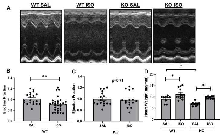
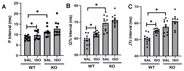
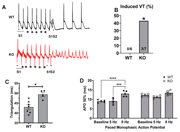
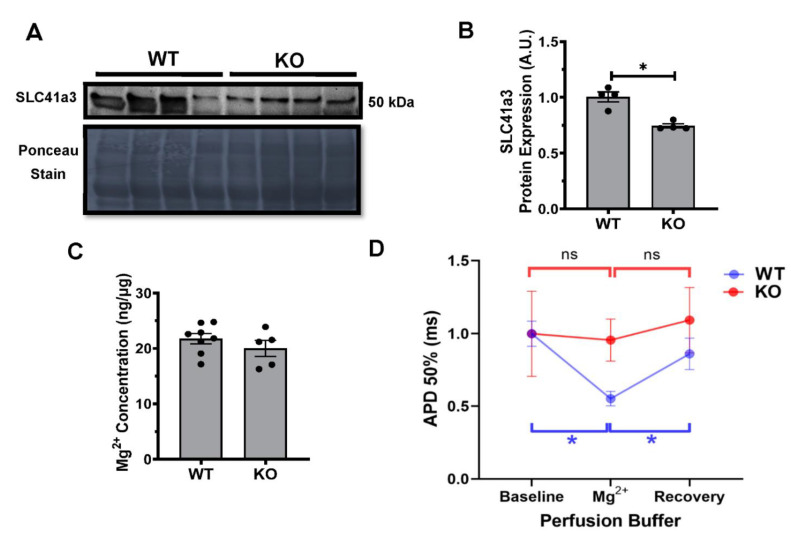
Kvβ subunits belong to the aldo-keto reductase superfamily, which plays a significant role in ion channel regulation and modulates the physiological responses. However, the role of Kvβ2 in cardiac pathophysiology was not studied, and therefore, in the present study, we hypothesized that Kvβ2 plays a significant role in cardiovascular pathophysiology by modulating the cardiac excitability and gene responses. We utilized an isoproterenol-infused mouse model to investigate the role of Kvβ2 and the cardiac function, biochemical changes, and molecular responses. The deletion of Kvβ2 attenuated the QTc (corrected QT interval) prolongation at the electrocardiographic (ECG) level after a 14-day isoproterenol infusion, whereas the QTc was significantly prolonged in the littermate wildtype group. Monophasic action potentials verified the ECG changes, suggesting that cardiac changes and responses due to isoproterenol infusion are mediated similarly at both the in vivo and ex vivo levels. Moreover, the echocardiographic function showed no further decrease in the ejection fraction in the isoproterenol-stimulated Kvβ2 knockout (KO) group, whereas the wildtype mice showed significantly decreased function. These experiments revealed that Kvβ2 plays a significant role in cardiovascular pathophysiology. Furthermore, the present study revealed SLC41a3, a major solute carrier transporter affected with a significantly decreased expression in KO vs. wildtype hearts. The electrical function showed that the decreased expression of SLC41a3 in Kvβ2 KO hearts led to decreased Mg2+ responses, whereas, in the wildtype hearts, Mg2+ caused action potential duration (APD) shortening. Based on the in vivo, ex vivo, and molecular evaluations, we identified that the deletion of Kvβ2 altered the cardiac pathophysiology mediated by SLC41a3 and altered the NAD (nicotinamide adenine dinucleotide)-dependent gene responses.
Keywords: Kvβ subunit; action potential; aldo-keto reductase; heart; magnesium; potassium channel; pyridine nucleotides; redox.
Conflict of interest statement
The authors declare no conflict of interest.
Figures


Similar articles
-
Genetic deletion of Kvβ2 (AKR6) causes loss of muscle function and increased inflammation in mice.Front Aging. 2023 Jun 12;4:1175510. doi: 10.3389/fragi.2023.1175510. eCollection 2023. Front Aging. 2023. PMID: 37377453 Free PMC article.
-
Kvβ1.1 (AKR6A8) senses pyridine nucleotide changes in the mouse heart and modulates cardiac electrical activity.Am J Physiol Heart Circ Physiol. 2017 Mar 1;312(3):H571-H583. doi: 10.1152/ajpheart.00281.2016. Epub 2016 Dec 16. Am J Physiol Heart Circ Physiol. 2017. PMID: 27986658 Free PMC article.
-
Metabolic regulation of Kv channels and cardiac repolarization by Kvβ2 subunits.J Mol Cell Cardiol. 2019 Dec;137:93-106. doi: 10.1016/j.yjmcc.2019.09.013. Epub 2019 Oct 19. J Mol Cell Cardiol. 2019. PMID: 31639389 Free PMC article.
-
Heteromeric complexes of aldo-keto reductase auxiliary KVβ subunits (AKR6A) regulate sarcolemmal localization of KV1.5 in coronary arterial myocytes.Chem Biol Interact. 2017 Oct 1;276:210-217. doi: 10.1016/j.cbi.2017.03.011. Epub 2017 Mar 22. Chem Biol Interact. 2017. PMID: 28342889 Free PMC article.
-
Biochemical and physiological properties of K+ channel-associated AKR6A (Kvβ) proteins.Chem Biol Interact. 2019 May 25;305:21-27. doi: 10.1016/j.cbi.2019.03.023. Epub 2019 Mar 26. Chem Biol Interact. 2019. PMID: 30926318 Free PMC article. Review.
Cited by
-
The Importance of Vitamin D and Magnesium in Athletes.Nutrients. 2025 May 13;17(10):1655. doi: 10.3390/nu17101655. Nutrients. 2025. PMID: 40431395 Free PMC article. Review.
-
Magnesium and Migraine.Nutrients. 2025 Feb 18;17(4):725. doi: 10.3390/nu17040725. Nutrients. 2025. PMID: 40005053 Free PMC article. Review.
-
NAD+ centric mechanisms and molecular determinants of skeletal muscle disease and aging.Mol Cell Biochem. 2022 Jun;477(6):1829-1848. doi: 10.1007/s11010-022-04408-1. Epub 2022 Mar 25. Mol Cell Biochem. 2022. PMID: 35334034 Free PMC article. Review.
-
Genetic deletion of Kvβ2 (AKR6) causes loss of muscle function and increased inflammation in mice.Front Aging. 2023 Jun 12;4:1175510. doi: 10.3389/fragi.2023.1175510. eCollection 2023. Front Aging. 2023. PMID: 37377453 Free PMC article.
References
-
- Tur J., Badole S.L., Cheng F., Das A., Kukreja R.C., Tipparaju S.M. Corticosteroids and aldose reductase inhibitor Epalrestat modulates cardiac action potential via Kvβ1.1 (AKR6A8) subunit of voltage-gated potassium channel. Mol. Cell. Biochem. 2017;436:71–78. doi: 10.1007/s11010-017-3079-9. - DOI - PMC - PubMed
Grants and funding
LinkOut - more resources
Full Text Sources
Other Literature Sources
Research Materials
