. 2021 Mar 29;22(7):3523.
doi: 10.3390/ijms22073523.

High Diversity of β-Glucosidase-Producing Bacteria and Their Genes Associated with Scleractinian Corals

Affiliations

High Diversity of β-Glucosidase-Producing Bacteria and Their Genes Associated with Scleractinian Corals

Hongfei Su et al. Int J Mol Sci. .

Abstract

β-Glucosidase is a microbial cellulose multienzyme that plays an important role in the regulation of the entire cellulose hydrolysis process, which is the rate-limiting step in bacterial carbon cycling in marine environments. Despite its importance in coral reefs, the diversity of β-glucosidase-producing bacteria, their genes, and enzymatic characteristics are poorly understood. In this study, 87 β-glucosidase-producing cultivable bacteria were screened from 6 genera of corals. The isolates were assigned to 21 genera, distributed among three groups: Proteobacteria, Firmicutes, and Actinobacteria. In addition, metagenomics was used to explore the genetic diversity of bacterial β-glucosidase enzymes associated with scleractinian corals, which revealed that these enzymes mainly belong to the glycosidase hydrolase family 3 (GH3). Finally, a novel recombinant β-glucosidase, referred to as Mg9373, encompassing 670 amino acids and a molecular mass of 75.2 kDa, was classified as a member of the GH3 family and successfully expressed and characterized. Mg9373 exhibited excellent tolerance to ethanol, NaCl, and glucose. Collectively, these results suggest that the diversity of β-glucosidase-producing bacteria and genes associated with scleractinian corals is high and novel, indicating great potential for applications in the food industry and agriculture.

Keywords: cultivable bacteria; diversity; metagenomic approach; scleractinian corals; β-glucosidase.


Conflict of interest statement

All authors edited and approved the manuscript. The authors declare no conflict of interest.

Figures
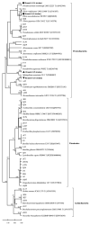
Figure 1
Figure 1
Phylogenetic tree of the cultivable β-glucosidase-producing bacteria isolated from scleractinian corals based on 16S rRNA gene sequences. The tree was constructed by the neighbor-joining method using MEGA package version 5.0. Only bootstrap values greater than 50% are presented in the nodes. The scale bar represents 2% nucleotide substitution. Branch 1 indicates nine Vibrio strains similar to Vibrio rotiferianus LMG21460T (AJ316187). Branch 2 indicates 13 Photobacterium strains similar to Photobacterium rosenbergii LMG 22223 T (AJ842344). Branch 3 indicates eight Sphingobium strains similar to Sphingobium wenxiniae JZ-1 T (FJ686047). Branch 4 indicates 11 Brevundimonas strains similar to Brevundimonas intermedia ATCC 15262 T (AJ227786). The circle indicated that 14 strains were from Favia, and the sequence similarity between them and Photobacterium rosenbergii was 99%, which were clustered into branch 1; The square indicates that a total of 9 strains isolated from Pocillopora, Porites, Acropora and Montipora have sequence similarity of 98.5–99.2% with Vibrio rotierianus, and clustered into branch 2; The trigonometry indicates that 9 strains with 97.5–99.1% similarity to Sphingobium wenxiniae JZ-1 clustered into branch 3; The inverted triangle indicated that the 11 strains were most similar to Paracoccus marcusii in similarity and clustered into branch 4. Pocillopora, Acropora, Porites, Favia, Turbinaria, and Montipora are represented by A, B, C, D, E, and F.
Figure 2
Figure 2
Carbohydrate-active enzymes: Auxiliary Activities (AA); Carbohydrate-Binding Modules (CBM); Carbohydrate Esterases (CE); Glycoside Hydrolases (GH); Glycosyl Transferases (GT); Polysaccharide Lyases (PL).
Figure 3
Figure 3
Amino acid sequence homologies of GH1 and GH3 β-glucosidase gene fragments from coral microbiology metagenomic DNA to known β-glucosidase.
Figure 4
Figure 4
Maximum Likelihood phylogenetic tree of the GH3 amino acid sequences. Amino acid sequences larger than 500 were selected from the GH3 family with the largest number for tree building analysis.
Figure 5
Figure 5
Molecular weight of Mg9373 determined by SDS-PAGE. M: protein molecular weight markers; 1: supernatant of cell lysis from E. coli BL21 DE3 cells; 2: supernatant of cell lysis from recombinant E. coli BL21 DE3 cells harboring pEASY-E1-mg9373 plasmid; 3: purified enzyme Mg9373 from Ni2+ column.
Figure 6
Figure 6
Effect of pH and temperature of Mg9373 on active and stability. (A) Effect of pH on active. The effect of pH on Mg9373 activity was determined at McIlvaine buffer between pH 3.0–8.0 and Glycine-NaOH buffer between pH 8.0–11.0, at 40 °C. (B) ffect of temperature on active and thermostability assay. The activity was determined in McIlvaine buffer (pH 7.0) at various temperatures (0–70 °C) and the thermostability of Mg9373 was determined after incubation at various temperatures (0–70 °C).
Figure 7
Figure 7
Effect of NaCl (A) and gluose (B) of Mg9373 on active and stability. (A) Effect of NaCl on stability. The stability of Mg9373 was determined at 40 °C in McIlvaine buffer (pH 6.0) after incubation at various NaCl (0–5 M). (B) Effect of glucose stability. The stability of Mg9373 was determined after incubation at various glucose (0–20%), at 40 °C in McIlvaine buffer (pH 6.0).
Figure 8
Figure 8
Michaelis-Menten plots for the reactions with pNPG. The inset shows the Lineweaver-Burk plots. Each data point represents the mean ± SD of three independent experiments.

References

    1. Forest R., Victor S., Farooq A., Nancy K. Diversity and distribution of coral-associated bacteria. Mar. Ecol. Prog. Ser. 2002;243:1–10.
    1. Pernice M., Raina J.B., Radecker N., Cardenas A., Pogoreutz C., Voolstra C.R. Down to the bone: The role of overlooked endolithic microbiomes in reef coral health. ISME J. 2020;14:325–334. doi: 10.1038/s41396-019-0548-z. - DOI - PMC - PubMed
    1. Eugene R. The role of microorganisms in coral health, disease and evolution. Nat. Rev. Microbiol. 2007;5:355–362. - PubMed
    1. Garren M., Azam F. New method for counting bacteria associated with coral mucus. Appl. Environ. Microbiol. 2010;76:6128–6133. doi: 10.1128/AEM.01100-10. - DOI - PMC - PubMed
    1. Garren M., Azam F. New directions in coral reef microbial ecology. Environ. Microbiol. 2012;14:833–844. doi: 10.1111/j.1462-2920.2011.02597.x. - DOI - PubMed

Substances

LinkOut - more resources