Review
. 2021 Mar 29;13(4):575.
doi: 10.3390/v13040575.

Transcriptional and Non-Transcriptional Activation, Posttranslational Modifications, and Antiviral Functions of Interferon Regulatory Factor 3 and Viral Antagonism by the SARS-Coronavirus

Affiliations
Review

Transcriptional and Non-Transcriptional Activation, Posttranslational Modifications, and Antiviral Functions of Interferon Regulatory Factor 3 and Viral Antagonism by the SARS-Coronavirus

Anna Glanz et al. Viruses. .

Abstract

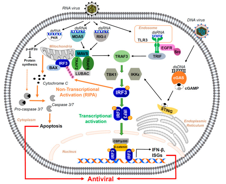
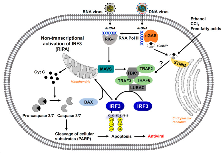
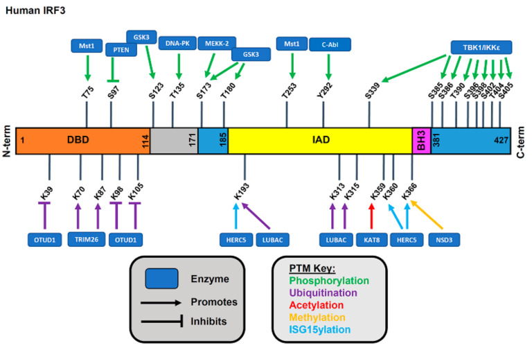
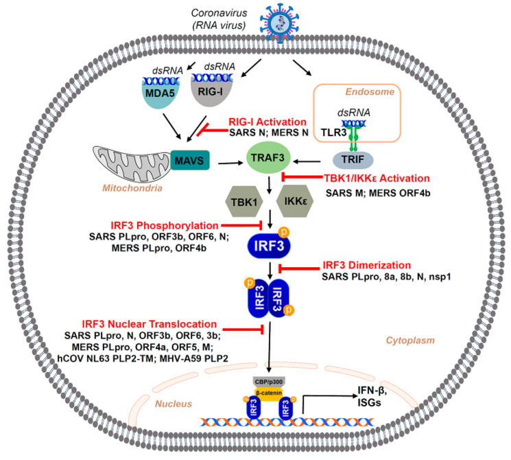
The immune system defends against invading pathogens through the rapid activation of innate immune signaling pathways. Interferon regulatory factor 3 (IRF3) is a key transcription factor activated in response to virus infection and is largely responsible for establishing an antiviral state in the infected host. Studies in Irf3-/- mice have demonstrated the absence of IRF3 imparts a high degree of susceptibility to a wide range of viral infections. Virus infection causes the activation of IRF3 to transcribe type-I interferon (e.g., IFNβ), which is responsible for inducing the interferon-stimulated genes (ISGs), which act at specific stages to limit virus replication. In addition to its transcriptional function, IRF3 is also activated to trigger apoptosis of virus-infected cells, as a mechanism to restrict virus spread within the host, in a pathway called RIG-I-like receptor-induced IRF3 mediated pathway of apoptosis (RIPA). These dual functions of IRF3 work in concert to mediate protective immunity against virus infection. These two pathways are activated differentially by the posttranslational modifications (PTMs) of IRF3. Moreover, PTMs regulate not only IRF3 activation and function, but also protein stability. Consequently, many viruses utilize viral proteins or hijack cellular enzymes to inhibit IRF3 functions. This review will describe the PTMs that regulate IRF3's RIPA and transcriptional activities and use coronavirus as a model virus capable of antagonizing IRF3-mediated innate immune responses. A thorough understanding of the cellular control of IRF3 and the mechanisms that viruses use to subvert this system is critical for developing novel therapies for virus-induced pathologies.

Keywords: IRF3; RIPA; SARS-CoV-2; innate antiviral immunity; interferon; posttranslational modifications; viral antagonism.


Conflict of interest statement

The authors declare no conflict of interest. The funders had no role in the design of the study, in the writing of the manuscript, or in the decision to publish the results.

Figures

Figure 1
Figure 1
The transcriptional and non-transcriptional activation of IRF3 by virus-activated signaling pathways lead to host antiviral response. The PRRs, e.g., TLR3, RIG-I, MDA-5, and cGAS, present at different cellular locations, recognize the viral nucleic acids (RNA or DNA) upon their entry into the host cell. In the transcriptional activation of IRF3, the PRR-mediated signaling pathways activate the downstream kinases, TBK1 and IKKε, and directly phosphorylate the cytosolic IRF3. The phosphorylated IRF3 translocates to the nucleus to transcribe the antiviral genes, e.g., IFNβ and ISG. In the non-transcriptional pathway, IRF3 gets activated by ubiquitination and translocates to the mitochondria to activate the intrinsic caspases, leading to apoptotic cell death. The transcriptional and non-transcriptional pathways of IRF3 contribute to the overall antiviral response of the host. A dsRNA-binding protein PKR can also trigger cellular apoptosis by activating eIF2α, which inhibits protein synthesis.
Figure 2
Figure 2
The non-transcriptional activation of IRF3 by RIPA causes cell death. IRF3 can be activated by RNA or DNA virus infection, which triggers RIG-I or cGAS signaling pathways. The RIG-I activation by RNA or DNA viruses can recruit a complex by TBK1/TRAF2/TRAF6, which allows the binding of IRF3 to LUBAC. The LUBAC-mediated linear ubiquitination of IRF3 leads to its translocation to the mitochondria and interaction with BAX. Mitochondrial translocation of IRF3/BAX complex leads to cytochrome C release that activates the apoptotic caspases, leading to apoptotic cell death. This pathway has been named ‘RIPA’, which can also be triggered by ethanol, CCL4, and free fatty acids, which activate the PRR signaling pathways.
Figure 3
Figure 3
Activation of IRF3 by posttranslational modifications. Human IRF3 and its various functional domains are shown; DBD, DNA-binding domain, IAD, IRF association domain, BH3, and Bcl2 homology domain. The amino acids that are posttranslationally modified are indicated, and their respective enzymes catalyze these modifications.
Figure 4
Figure 4
SARS-coronavirus (SARS-CoV) mediated inhibition of IRF3 activation. The SARS-CoV has various proteins that play a role in antagonizing IRF3 activation as a defense mechanism. Its nucleocapsid (N protein) acts by inhibiting RIG-I activation, one of the major PRRs responsible for IRF3 activation. Matrix (M) protein of SARS inhibits the activation of kinases (TBK-1, IKKε) required to activate IRF3. Various accessory proteins (Orf 3b, 6), protease (PLpro), as well as the N protein of the virus can antagonize the phosphorylation and activation of IRF3 itself. Even after IRF3 activation, SARS-CoV can use several accessory proteins (8a, 8b, 3b, 6) and non-structural proteins (nsp1) or the N-protein to block major changes, such as IRF3 dimerization or its nuclear translocation, that are essential for IRF3 to transcribe its downstream antiviral genes successfully. Some of these activation steps are also inhibited by MERS-CoV proteins, as shown in the cartoon.

Similar articles

Cited by

References

    1. Marsili G., Perrotti E., Remoli A.L., Acchioni C., Sgarbanti M., Battistini A. IFN Regulatory Factors and Antiviral Innate Immunity: How Viruses Can Get Better. J. Interferon Cytokine Res. 2016;36:414–432. doi: 10.1089/jir.2016.0002. - DOI - PubMed
    1. Jefferies C.A. Regulating IRFs in IFN Driven Disease. Front. Immunol. 2019;10:325. doi: 10.3389/fimmu.2019.00325. - DOI - PMC - PubMed
    1. Ozato K., Tailor P., Kubota T. The interferon regulatory factor family in host defense: Mechanism of action. J. Biol. Chem. 2007;282:20065–20069. doi: 10.1074/jbc.R700003200. - DOI - PubMed
    1. Hiscott J. Triggering the innate antiviral response through IRF-3 activation. J. Biol. Chem. 2007;282:15325–15329. doi: 10.1074/jbc.R700002200. - DOI - PubMed
    1. Petro T.M. IFN Regulatory Factor 3 in Health and Disease. J. Immunol. 2020;205:1981–1989. doi: 10.4049/jimmunol.2000462. - DOI - PubMed

Publication types

Substances

LinkOut - more resources