Review
. 2021 Mar 29;12(4):505.
doi: 10.3390/genes12040505.

LRRK2 at the Crossroad of Aging and Parkinson's Disease

Affiliations
Review

LRRK2 at the Crossroad of Aging and Parkinson's Disease

Eun-Mi Hur et al. Genes (Basel). .

Abstract

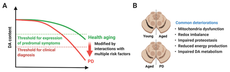
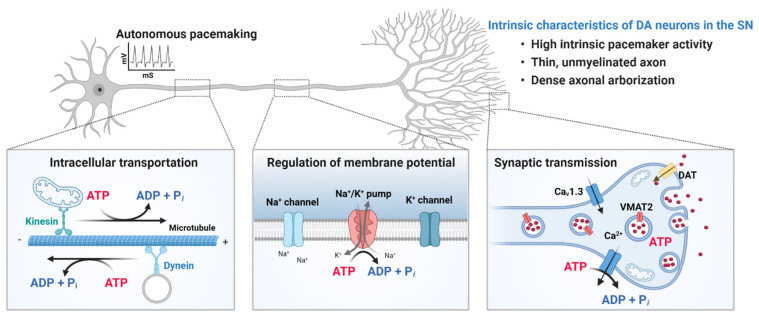
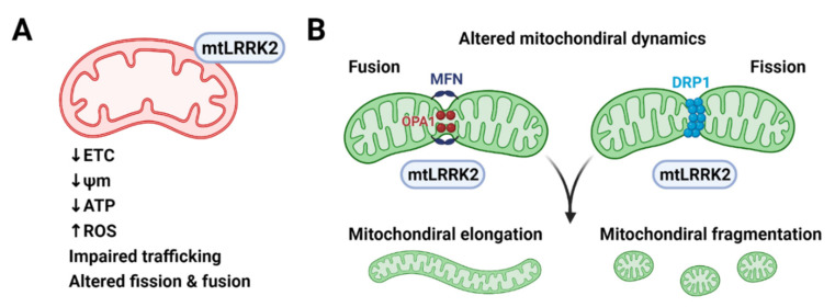
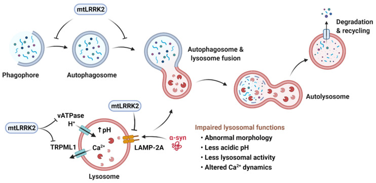
Parkinson's disease (PD) is a heterogeneous neurodegenerative disease characterized by the progressive loss of dopaminergic neurons in the substantia nigra pars compacta and the widespread occurrence of proteinaceous inclusions known as Lewy bodies and Lewy neurites. The etiology of PD is still far from clear, but aging has been considered as the highest risk factor influencing the clinical presentations and the progression of PD. Accumulating evidence suggests that aging and PD induce common changes in multiple cellular functions, including redox imbalance, mitochondria dysfunction, and impaired proteostasis. Age-dependent deteriorations in cellular dysfunction may predispose individuals to PD, and cellular damages caused by genetic and/or environmental risk factors of PD may be exaggerated by aging. Mutations in the LRRK2 gene cause late-onset, autosomal dominant PD and comprise the most common genetic causes of both familial and sporadic PD. LRRK2-linked PD patients show clinical and pathological features indistinguishable from idiopathic PD patients. Here, we review cellular dysfunctions shared by aging and PD-associated LRRK2 mutations and discuss how the interplay between the two might play a role in PD pathologies.

Keywords: LRRK2; Parkinson’s disease; ROS; aging; autophagy; lysosome; mitochondria.


Conflict of interest statement

The authors declare no conflict of interest.

Figures

Figure 1
Figure 1
A hypothetical model of aging and Parkinson’s disease (PD). (A) Functional DA neurons are reduced in both healthy aging and PD. Rate of degeneration in healthy aging and PD might be similar up to a certain time point or age, but degeneration in PD is accelerated until reaching the threshold for clinical diagnosis (red dotted line). The upper green dotted line represents the threshold for expression of prodromal symptoms. Several genetic and environmental risk factors might play a role in accelerating degeneration of DA neurons and causing PD. (B) Degeneration of DA neurons in the SN is considered as the pathological hallmark of PD, but it also occurs in healthy aging. The difference in healthy aging and PD might be the quantity of neurodegeneration rather than the quality. Healthy aging and PD share a plethora of cellular dysfunctions, such as mitochondrial dysfunction, redox imbalance, impaired proteostasis, reduced energy production, and impaired DA metabolism. Created with BioRender.com.
Figure 2
Figure 2
Intrinsic properties of dopaminergic (DA) neurons in the substantia nigra (SN). The nigrostriatal DA neurons are autonomous pacemakers, and their tonic spontaneous activity is important for the sustained release of dopamine in target structures, such as the striatum. In addition, DA neurons in the SN have thin, unmyelinated axons with an extraordinary large axonal arbor and a very high number of synaptic terminals. The size and complexity of DA neurons in the SN impose a high metabolic burden to produce ATP, which is required for transporting cellular components to appropriate locations, maintaining resting membrane potential, generating and propagating action potential, and regulating synaptic transmission. Metabolically active DA neurons in the SN are susceptible to mitochondrial dysfunction, which is a prominent feature of both aging and PD. Created with BioRender.com.
Figure 3
Figure 3
Mitochondrial dysfunction caused by pathogenic LRRK2 mutants. (A) Pathogenic LRRK2 mutants (mtLRRK2) reduce the activity of electron transfer chain (ETC) reaction and mitochondria membrane potential (ψm), resulting in inefficient ATP production and increased reactive oxygen species (ROS) production. (B) Pathogenic LRRK2 mutants interact with mitochondria fusion proteins, such as mitofusin (MFN) and optic atrophy protein 1 (OPA1) and mitochondria fission proteins, such as dynamin-related protein 1 (DRP1). Pathogenic LRRK2 mutants alter mitochondrial morphology and dynamics presumably by interacting with the fusion and fission machinery but the exact molecular mechanism awaits to be elucidated. Created with BioRender.com (accessed on 13 February 2021).
Figure 4
Figure 4
Pathogenic LRRK2 mutants (mtLRRK2) and the autophagy-lysosomal pathways. Macroautophagy is initiated by the formation of a cup-shaped membrane, termed phagophore, which engulfs damaged organelles or misfolded proteins to form the autophagosome. The autophagosome fuses with the lysosome and generates the autolysosome, in which the contents are degraded. Transition into autophagosome and autolysosome is inhibited by mtLRRK2. In chaperone-mediated autophagy, proteins are targeted to lysosomes by a chaperone through interaction between the chaperone and a pentapeptide present within the substrate. Substrate proteins then bind to a transmembrane receptor, lysosome-associated membrane protein type 2A (LAMP-2A), which multimerizes to form the translocation complex that carries the substrate proteins into the lysosome for degradation. LAMP-2A is inhibited by mtLRRK2, leading to the accumulation of CMA substrates, such as α-synuclein (α-syn). mtLRRK2 can also decrease lysosomal acidity and disrupt lysosomal calcium dynamics by inhibiting lysosomal H+-ATPase pump, vATPase and TRPML1. Created with BioRender.com.

References

    1. Wakabayashi K., Tanji K., Odagiri S., Miki Y., Mori F., Takahashi H. The Lewy body in Parkinson’s disease and related neurodegenerative disorders. Mol. Neurobiol. 2013;47:495–508. doi: 10.1007/s12035-012-8280-y. - DOI - PubMed
    1. Kouli A., Torsney K.M., Kuan W.L. Parkinson’s Disease: Etiology, Neuropathology, and Pathogenesis. In: Stoker T.B., Greenland J.C., editors. Parkinson’s Disease: Pathogenesis and Clinical Aspects. Exon Publications; Brisbane, QLD, Australia: 2018. - DOI - PubMed
    1. Warner T.T., Schapira A.H. Genetic and environmental factors in the cause of Parkinson’s disease. Ann. Neurol. 2003;53(Suppl. S3):S16–S23; discussion S23–S15. doi: 10.1002/ana.10487. - DOI - PubMed
    1. Reeve A., Simcox E., Turnbull D. Ageing and Parkinson’s disease: Why is advancing age the biggest risk factor? Ageing Res. Rev. 2014;14:19–30. doi: 10.1016/j.arr.2014.01.004. - DOI - PMC - PubMed
    1. Rodriguez M., Rodriguez-Sabate C., Morales I., Sanchez A., Sabate M. Parkinson’s disease as a result of aging. Aging Cell. 2015;14:293–308. doi: 10.1111/acel.12312. - DOI - PMC - PubMed

Publication types

Substances

LinkOut - more resources