. 2021 Mar 29;13(4):1126.
doi: 10.3390/nu13041126.

Uptake of Vitamins D2, D3, D4, D5, D6, and D7 Solubilized in Mixed Micelles by Human Intestinal Cells, Caco-2, an Enhancing Effect of Lysophosphatidylcholine on the Cellular Uptake, and Estimation of Vitamins D' Biological Activities

Affiliations

Uptake of Vitamins D2, D3, D4, D5, D6, and D7 Solubilized in Mixed Micelles by Human Intestinal Cells, Caco-2, an Enhancing Effect of Lysophosphatidylcholine on the Cellular Uptake, and Estimation of Vitamins D' Biological Activities

Eiichi Kotake-Nara et al. Nutrients. .

Abstract

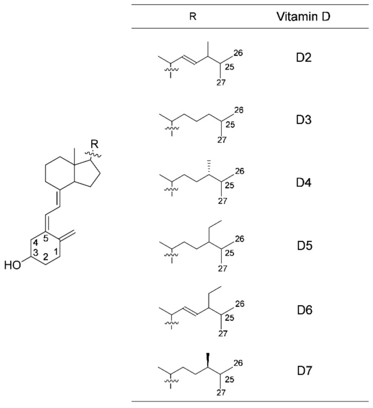
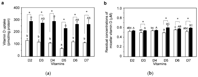
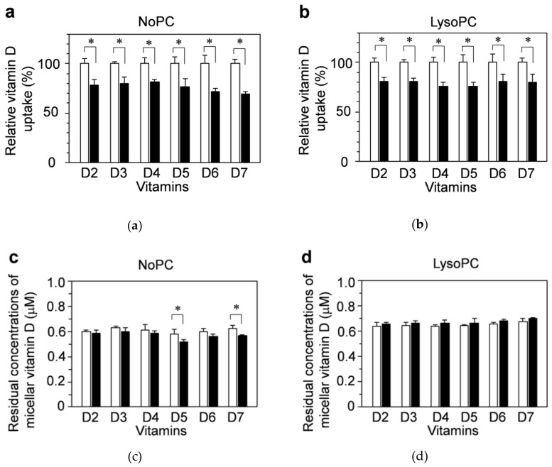
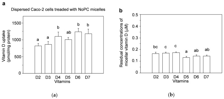
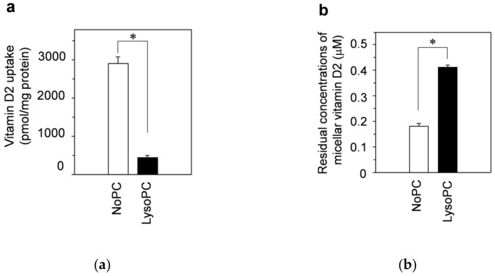
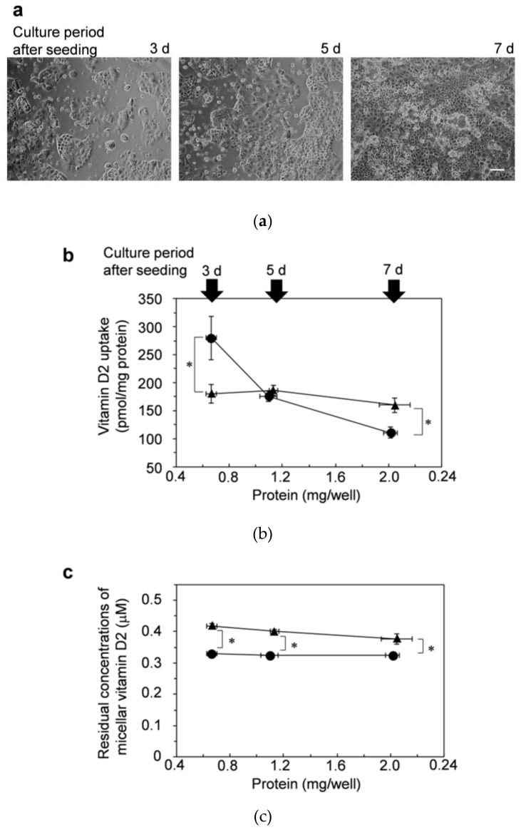
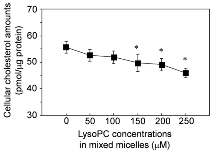
Vitamins D have various biological activities, as well as intestinal calcium absorption. There has been recent concern about insufficient vitamin D intake. In addition to vitamins D2 and D3, there are lesser-known vitamins D4-D7. We synthesized vitamins D5-D7, which are not commercially available, and then evaluated and compared the mixed micelles-solubilized vitamins D uptake by Caco-2 cells. Except for vitamin D5, the uptake amounts of vitamins D4-D7 by differentiated Caco-2 cells were similar to those of vitamins D2 and D3. The facilitative diffusion rate in the ezetimibe inhibited pathway was approximately 20% for each vitamin D type, suggesting that they would pass through the pathway at a similar rate. Lysophosphatidylcholine enhanced each vitamin D uptake by approximately 2.5-fold. Lysophosphatidylcholine showed an enhancing effect on vitamin D uptake by reducing the intercellular barrier formation of Caco-2 cells by reducing cellular cholesterol, suggesting that increasing the uptakes of vitamins D and/or co-ingesting them with lysophosphatidylcholine, would improve vitamin D insufficiency. The various biological activities in the activated form of vitamins D4-D7 were estimated by Prediction of Activity Spectra for Substances (PASS) online simulation. These may have some biological activities, supporting the potential as nutritional components.

Keywords: Caco-2 cells; PASS; intercellular barrier formation; intestinal uptake; lysophosphatidylcholine; mixed micelles; vitamin D.


Conflict of interest statement

The authors declare no conflict of interest.

Figures

Figure 1
Figure 1
Chemical structures of vitamins D. The numbers in the structural formula indicate the locations of the carbons.
Figure 2
Figure 2
Uptake of vitamin D solubilized in mixed micelles by differentiated Caco-2 cells and the effects of lysophosphatidylcholine. Differentiated Caco-2 cells were incubated for 2 h with vitamin D in mixed micelles without lysophosphatidylcholine and mixed micelles with lysophosphatidylcholine, called NoPC mixed micelles (open bars) and lysoPC mixed micelles (filled bars), respectively. (a) Vitamin D uptake by differentiated Caco-2 cells. (b) Residual concentrations of vitamin D in the mixed micelles (micellar vitamin D) after 2-h incubation with the cells. Data are presented as the means ± standard deviation of four wells in a single experiment. Replicate experiments showed a similar trend. Statistical analyses were performed between NoPC mixed micelle and lysoPC mixed micelle in same vitamin D, among 6 kinds of vitamins D in NoPC mixed micelles, and among 6 kinds of vitamins D in lysoPC mixed micelles in each graph. The asterisk indicates a value with significant difference as determined by unpaired t-test (p < 0.05). Values not sharing a common letter were significantly different as determined by Tukey–Kramer test (p < 0.05).
Figure 3
Figure 3
Inhibitory effects of ezetimibe on the uptake of vitamin D solubilized in mixed micelles by differentiated Caco-2 cells. Differentiated Caco-2 cells were pretreated for 2 h with the vehicle (1.5% dimethyl sulfoxide (DMSO), open bars) alone or with 150 μM ezetimibe (filled bars), which is an inhibitor of the protein Niemann-Pick C1-like 1 (NPC1L1), and then incubated for 2 h with vitamin D in NoPC mixed micelles (left side) and lysoPC mixed micelles (right side). (a,b): Vitamin D uptake by differentiated Caco-2 cells. (c,d): Residual concentrations of micellar vitamin D after 2-h incubation with the cells. Data are presented as the means ± standard deviation of four wells in a single experiment. Replicate experiments showed a similar trend. Statistical analysis was performed to compare each vitamin D with and without ezetimibe. The asterisk indicates a value with significant difference as determined by unpaired t-test (p < 0.05).
Figure 4
Figure 4
Uptake of vitamin D solubilized in mixed micelles by dispersed Caco-2 cells. Dispersed Caco-2 cells without cell-cell adhesion/cell-matrix adhesion were incubated for 2 h with vitamin D in NoPC mixed micelles. The cells were prepared by dispersing the cells with trypsin. (a) Vitamin D uptake by dispersed Caco-2 cells. (b) Residual concentrations of the micellar vitamin D after 2-h incubation with the cells. Data are presented as the means ± standard deviation of four wells in a single experiment. Replicate experiments showed a similar trend. Statistical analysis was performed among all six samples in each graph. Values not sharing a common letter were significantly different as determined by Tukey–Kramer test (p < 0.05).
Figure 5
Figure 5
Effects of lysophosphatidylcholine on the uptake of vitamin D2 solubilized in mixed micelles by dispersed Caco-2 cells. Dispersed Caco-2 cells without cell-cell adhesion/cell-matrix adhesion were incubated for 2 h with vitamin D2 in NoPC mixed micelles and lysoPC mixed micelles. (a) Vitamin-D2 uptake by dispersed Caco-2 cells. (b) Residual concentrations of micellar vitamin D2 after 2-h incubation with the cells. Data are presented as the means ± standard deviation of four wells in a single experiment. Replicate experiments showed a similar trend. Statistical analysis was performed among all six samples in each graph. The asterisks indicate significant differences as determined by unpaired t-test (p < 0.05).
Figure 6
Figure 6
Effects of lysophosphatidylcholine on the uptake of vitamin D2 by Caco-2 cells with insufficient cell-cell adhesion. Caco-2 cells with insufficient cell-cell adhesion/cell-matrix adhesion were prepared by cultivating for 3, 5, and 7 d after seeding in the culture plate. (a) These cells were observed by phase-contrast microscopy (100×). The white line in the photo of 7 d indicates the scale bar (100 μm). The adhered Caco-2 cells were then incubated for 2 h with vitamin D2 in NoPC mixed micelles (circles) and lysoPC mixed micelles (triangles). (b) Vitamin-D2 uptake by the adherent Caco-2 cells. (c) Residual concentrations of the micellar vitamin D2 after 2-h incubation with the cells. Data are presented as the means ± standard deviation of four wells in a single experiment. Replicate experiments showed a similar trend. Statistical analysis was performed between two mixed micelles at the same cultivation time (3, 5, and 7 d). The asterisk indicates a value with significant difference as determined by unpaired t-test (p < 0.05).
Figure 7
Figure 7
Reducing effect of lysophosphatidylcholine in mixed micelles on cellular cholesterol amounts in the differentiated Caco-2 cells. The differentiated Caco-2 cells were incubated for 2 h with the mixed micelles containing lysophosphatidylcholine at 0–250 μM. The mixed micelles contained no vitamin D. Data are presented as the means ± standard deviation of four wells in a single experiment. Replicate experiments showed a similar trend. The asterisks indicate significant differences from the value of lysophosphatidylcholine at 0 μM as determined by Dunnett’s test (p < 0.05).

Similar articles

Cited by

References

    1. Miyauchi M., Hirai C., Nakajima H. The solar exposure time required for vitamin D3 synthesis in the human body estimated by numerical simulation and observation in Japan. J. Nutr. Sci. Vitaminol. 2013;59:257–263. doi: 10.3177/jnsv.59.257. - DOI - PubMed
    1. Godar D.E., Pope S.J., Grant W.B., Holick M.F. Solar UV doses of young Americans and vitamin D3 production. Environ. Health Perspect. 2012;120:139–143. doi: 10.1289/ehp.1003195. - DOI - PMC - PubMed
    1. MacLaughlin J., Holick M.F. Aging decreases the capacity of human skin to produce vitamin D3. J. Clin. Investig. 1985;76:1536–1538. doi: 10.1172/JCI112134. - DOI - PMC - PubMed
    1. Afzal S., Nordestgaard B.G., Bojesen S.E. Plasma 25-hydroxyvitamin D and risk of non-melanoma and melanoma skin cancer: A prospective cohort study. J. Investig. Dermatol. 2013;133:629–636. doi: 10.1038/jid.2012.395. - DOI - PubMed
    1. Lee D.U., Weon K.Y., Nam D.Y., Nam J.H., Kim W.K. Skin protective effect of guava leaves against UV-induced melanogenesis via inhibition of ORAI1 channel and tyrosinase activity. Exp. Dermatol. 2016;25:977–982. doi: 10.1111/exd.13151. - DOI - PubMed

LinkOut - more resources