Review
. 2021 Mar 29;22(7):3553.
doi: 10.3390/ijms22073553.

Skeletal Aging and Osteoporosis: Mechanisms and Therapeutics

Affiliations
Review

Skeletal Aging and Osteoporosis: Mechanisms and Therapeutics

Abhishek Chandra et al. Int J Mol Sci. .

Abstract

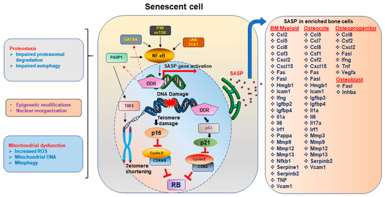
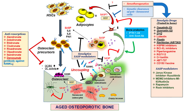
Bone is a dynamic organ maintained by tightly regulated mechanisms. With old age, bone homeostasis, which is maintained by an intricate balance between bone formation and bone resorption, undergoes deregulation. Oxidative stress-induced DNA damage, cellular apoptosis, and cellular senescence are all responsible for this tissue dysfunction and the imbalance in the bone homeostasis. These cellular mechanisms have become a target for therapeutics to treat age-related osteoporosis. Genetic mouse models have shown the importance of senescent cell clearance in alleviating age-related osteoporosis. Furthermore, we and others have shown that targeting cellular senescence pharmacologically was an effective tool to alleviate age- and radiation-induced osteoporosis. Senescent cells also have an altered secretome known as the senescence associated secretory phenotype (SASP), which may have autocrine, paracrine, or endocrine function. The current review discusses the current and potential pathways which lead to a senescence profile in an aged skeleton and how bone homeostasis is affected during age-related osteoporosis. The review has also discussed existing therapeutics for the treatment of osteoporosis and rationalizes for novel therapeutic options based on cellular senescence and the SASP as an underlying pathogenesis of an aging bone.

Keywords: SASP; aging; osteoporosis; radiation; senescence; senotherapeutic.


Conflict of interest statement

The authors declare no conflict of interest.

Figures

Figure 1
Figure 1
Spectrum of changes in a senescent cell. DNA damage response (DDR) is one of the key inducers of cellular senescence, and if the DNA damage is in the telomere sites, this drives the cell towards a senescent state which has several characteristics, also acting as sustainers or inducers of the senescent state of the cell. Telomere shortening or damage driven DDR initiates the p16Ink4a or p21 driven pathways which block the cyclin D, cyclin dependent kinase (CDK)2/4/6, and cyclin E to thereby stabilizing the retinoblastoma (Rb) protein, allowing the cell to enter the arrest phase. Activation of nuclear factor kappa B (NF-кB) through indirect activation of PARP1, GATA4, p38/ mechanistic target of rapamycin (MTOR), or Janus Kinase/Signal Transducer and Activator of Transcription (JAK/STAT) pathways activate the transcription of senescence associated secretory phenotype (SASP) genes. Proteostasis, either by impairment in the ubiquitin proteasome system or the autophagy pathway, allows aggregation of unwanted proteins, contributing to senescent profile of the cell. Mitochondrial dysfunction, including changes in the mitochondrial DNA, increased reactive oxygen species (ROS) and altered autophagy of the mitochondrial compartments, contributing to the overall stressed environment leading to senescence. Chemokines, interleukins, and matrix modifying enzymes form the bulk of the proinflammatory SASP genes which may work in an autocrine, paracrine, or endocrine manner. The list of SASP proteins was generated based on their expression in enriched bone cells. SASP abbreviations—TRF2: telomeric repeat binding factor (TRF), Ccl: C-C motif chemokine ligand, Csf: colony-stimulating factor, Cxcl: chemokine (C-X-C motif) ligand, HMGB1: high mobility group box 1, Icam1: intercellular adhesion molecule 1, Ifng: interferon gamma, Igfbp: Insulin-like growth factor binding proteins, Il: Interleukin, Irf1: Interferon Regulatory Factor 1, Mmp: Matrix metallopeptidase, Pappa: pregnancy-associated plasma protein A, TNF: Tumor necrosis factor, Vcam1: Vascular Cell Adhesion Molecule 1.
Figure 2
Figure 2
Mechanisms underlying an aging skeleton and potential therapeutic options. Bone formation which entails recruitment of bone marrow stem cells (BMSCs) to the bone surface, differentiation into osteoblasts and mineralization by the osteoblasts is followed by further differentiation of osteoblasts into osteocytes, which embed in the matrix, thereby communicating with other osteocytes, or cells in the bone environment through canalicular networks. Hematopoietic stem cells (HSCs) and precursors to the osteoclasts are activated by the binding of the RANKL to the RANK receptor, promoting osteoclastogenesis and bone resorption. Osteoprotegerin (OPG) a decoy receptor to RANKL, secreted by the osteoblasts, blocks the binding of RANKL to RANK and blocks osteoclastogenesis. With aging, osteoblasts and osteocytes undergo apoptosis or cellular senescence, and in the process, this internal regulation by OPG is disturbed, leading to more resorption. Production of pro-inflammatory SASP exacerbates the suppression of osteoblast function while triggering an activation of osteoclast precursors towards osteoclastogenesis. Moreover, reduction in BMSCs due to an altered fate to adipogenesis, also contributes to the suppression of osteoblast function. Reduction in osteoclast numbers, but increased activity, also disturbs the recruitment of more BMSCs to the bone surface, thus causing uncoupling of the bone homeostasis. Bone anabolics such as PTH 1-34 and neutralizing antibody against sclerostin (Sost), and anti-resorptives as shown in the figure have been effective treatments for post-menopausal osteoporosis, but their efficacy in an aging population is not determined. Genetic removal of senescent cells was shown to restore bone homeostasis in aged mice hence pharmacological targeting senescent cells became a lucrative therapeutic option. Drugs that can remove the senescent cell (Senolytic drugs) or suppress the production of SASP (SASP modulators), collectively called senotherapeutics, may remove the triggers for uncoupling and restore bone homeostasis. Several of these senotherapeutics are listed in the figure and the ones which have been tested in some form of skeletal aging are underlined. Abbreviations- OC: osteoclast, BS: bone surface, Il: interleukin, PAI1: plasminogen activator inhibitor 1.

References

    1. Sozen T., Ozisik L., Basaran N.C. An overview and management of osteoporosis. Eur. J. Rheumatol. 2017;4:46–56. doi: 10.5152/eurjrheum.2016.048. - DOI - PMC - PubMed
    1. Manolagas S.C. From estrogen-centric to aging and oxidative stress: A revised perspective of the pathogenesis of osteoporosis. Endocr. Rev. 2010;31:266–300. doi: 10.1210/er.2009-0024. - DOI - PMC - PubMed
    1. Riggs B.L., Khosla S., Melton L.J., 3rd A unitary model for involutional osteoporosis: Estrogen deficiency causes both type I and type II osteoporosis in postmenopausal women and contributes to bone loss in aging men. J. Bone Miner. Res. 1998;13:763–773. doi: 10.1359/jbmr.1998.13.5.763. - DOI - PubMed
    1. Farr J.N., Rowsey J.L., Eckhardt B.A., Thicke B.S., Fraser D.G., Tchkonia T., Kirkland J.L., Monroe D.G., Khosla S. Independent Roles of Estrogen Deficiency and Cellular Senescence in the Pathogenesis of Osteoporosis: Evidence in Young Adult Mice and Older Humans. J. Bone Miner. Res. 2019;34:1407–1418. doi: 10.1002/jbmr.3729. - DOI - PMC - PubMed
    1. Khosla S., Pacifici R. Chapter 46–Estrogen Deficiency, Postmenopausal Osteoporosis, and Age-Related Bone Loss. In: Marcus R., Feldman D., Dempster D.W., Luckey M., Cauley J.A., editors. Osteoporosis. 4st ed. Academic Press; San Diego, CA, USA: 2013. pp. 1113–1136.

Substances

LinkOut - more resources